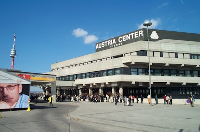
At the close of this year's European Congress of Radiology in Vienna, incoming ECR president Helen Carty from the U.K. spoke of a meeting based on friendship, a congress in which nations of "many creeds and races, rich and poor," come together to share their knowledge in a nonconfrontational atmosphere.
And so it seemed to go for the first all-digital edition of this increasingly global radiology meeting. The ECR gains attendees every year from, at last count, 56 nations. Professional attendance was up in 2003 by more than 13% compared to last year (7,614 vs. 6,722 in 2002), according to the latest count on the ECR's Web site (www.ecr.org).
Vendor attendance reached 4,814 this year, and the somewhat limited exhibition space was sold out long in advance. In scientific papers, ECR organizers dealt with this year's 21% increase in abstracts (1,570 submitted in 2003) by rejecting more than a third of them.
 |
|
This year’s ECR meeting saw professional attendance climb by 13%.
|
This year's new initiatives included the EPOS electronic poster system, which replaced traditional poster presentations at the meeting. EPOS workstations remained packed throughout the conference, with more than 10,000 presentations e-mailed to others in the five days of the meeting. The E3 (Excellence in Education in Europe) educational initiative, an ambitious scheme aimed at improving, modernizing, and standardizing radiology education throughout Europe, also got off to a good start.
Led by 2003 ECR president Dr. Nicholas Gourtsoyiannis from the University of Crete, this year's conference highlighted Greece and Turkey as featured nations in the new "ECR Meets" initiative. Greece and Turkey presented special sessions in ultrafast MR and interventional neuroradiology, respectively. Next year England and Ireland, along with Poland, will share the honors.
For next year's conference, to be held March 5-9, 2004 in Vienna, look for more interactive E3 educational offerings, new hands-on workshops in musculoskeletal radiology, ultrasound, and vertebroplasty, and a new foundation course in radiology with a focus on the abdomen.
Research
The specificity of computed tomography for lung cancer screening can be improved by identifying which patients actually need to be followed up, according to the results of a study of 3,000 screening subjects presented by Dr. Claudia Henschke of the Weill Medical College at Cornell University in New York City. No malignancies were found in baseline scans for any lesions smaller than 5 mm, she said, concluding that there is generally no need to follow up small lesions unless they appear in subsequent scans.
Cardiac imaging and coronary calcium assessment were also major themes in CT imaging. Dr. Bernd Wintersperger from the University of Munich in Germany led a study that found that a low-dose protocol for coronary artery calcium screening produces essentially the same results as standard-dose screening. In the study, 48 consecutive patients agreed to undergo coronary calcium screening twice. One protocol used 120 kVp and 133 mAs, the other 80 kVp with 300 mAs.
According to the results, the low-dose protocol showed significantly lower patient radiation exposure than the standard protocol (0.72 mSv versus 2.4 mSv, p < 0.0001). CT dose index (CTDI) phantom measurements showed a 65% dose reduction in the low-dose protocol: 23.45 vs. 10.11 for the low-dose exam. Moreover, the calcium scoring results were nearly identical (r=0.99, p < 0.0001). Algorithmic analysis also showed a very good correlation, with no significant differences between the 80-kVp protocol and the 120-kVp protocol.
In a potentially groundbreaking virtual colonoscopy study, researchers from the University of Muenster in Germany found that lowered radiation dose did not inhibit the detection of colorectal polyps, but thicker slices did.
Dr. Johann Wessling tested a full range of acquisition protocols in a water-filled acrylic phantom equipped with "polyps" ranging from 2 to 12 mm in size. According to the results, higher mean slice thicknesses produced blurred polyp borders and artificial enlargement of polyps. As for sensitivity, polyps 8 mm and larger were detected independently of slice thickness. Slice thickness of 1.25 mm was best for the detection of polyps overall, and was unaffected by tube-current reductions down to 10 mAs, he concluded.
For patients with advanced rectal carcinoma, MR was superior to both CT and ultrasound for determining the extent of local tumor invasion, according to a study by Dr. Jens Stollfus from the Technical University of Munich in Germany. In the study, T1 and T2 MR imaging, multislice CT, and ultrasound were performed in 103 patients with initial presentation of tumors T3 and greater.
MRI had a sensitivity of 84% for the detection of tumor invasion, compared with 69% and 71% for CT and ultrasound, respectively. In specificity, MRI achieved 68%, compared with 55% for CT and 28% for ultrasound.
MRI’s positive predictive value was 77%, compared with 65% for CT and 61% for US. The negative predictive value was 77% for MRI and 41% for US. However, CT tended to be more specific regarding the prediction of nodal status (p < 0.05). And the group found no significant difference between contrast-enhanced and unenhanced MRI for the detection of local tumor invasion.
In a comparative study with potentially important implications for myocardial assessment strategies, MRI outperformed PET in predicting myocardial recovery following coronary artery bypass grafting (CABG). Dr. Peter Hunold from University Hospital in Essen, Germany, led a study of 12 patients who underwent both PET and MRI before and after the revascularization procedure. According to the results, 71/143 segments predicted as viable by PET did not improve after CABG. Based on the identification of viable myocardium with functional recovery after revascularization, MRI had a sensitivity and specificity of 95% and 72%, respectively, compared with 97% and 47%, respectively, for PET.
For PET and CT as a team, however, two modalities were definitely better than one. In another study from the University of Essen, Germany, Dr. Gerald Antoch compared PET-CT to MRI for visualizing tumors in 50 patients. The modalities were found to be comparable in 33 (66%) of patients, notably in the abdominal organs.
PET-CT beat MRI in detecting lymph node and pulmonary lesions, however. In eight patients (16%) with cervical, thoracic, or abdominal lymph node tumors, PET-CT was able to detect tumors missed on MRI due to their small size (less than 1 cm). PET-CT spotted 94 pulmonary lesions in 16 patients, compared to 58 lesions in 13 patients for MRI. MRI excelled in visualizing bone metastases, however, finding metastases in seven patients (14%) that were missed on PET-CT.
"Whole-body PET-CT seems to be superior to whole-body MRI in detecting lymph node metastases and pulmonary lesions," Antoch said. "On the other hand, whole-body MRI detects bone metastases more accurately. Whole-body PET-CT and MR imaging complement each other in the staging of malignant diseases."
In another PET-CT presentation, Antoch and colleagues examined the utility of the two modalities in monitoring radiation therapy in the liver tissues of nine pig models. Following central bile duct resection and biliodigestive anastomosis, PET-CT liver scans were then conducted at two, four, and eight weeks to assess the impact of the irradiation on liver function as measured by glucose uptake.
Antoch concluded that PET is well suited for delineation of functional alterations following radiation therapy, while CT is appropriate for visualizing surgical alterations, vascular differences, and liver volume. He recommended first assessing an irradiated area with PET-CT, and then following up with PET and CT independently.
In a third PET-CT study, the Essen group tested a new breathing protocol it developed in which patients were asked to limit their breathing during crucial parts of image acquisition. The patients began by breathing quietly, and as the scan's field of view reached the mediastinum, the patients were told to exhale and hold their breath for the next 15 seconds -- until the scanning slice plane reached the lower end of the liver.
The group found that without using the protocol, there were displacements of up to 25 mm between the CT and PET images in the diaphragm area, the area most affected by respiration-related movement. According to the results, there were 25% more artifact-free cases when using the breathhold protocol in single-slice mode, and 50% more artifact-free cases when the PET-CT scanner was used in dual-slice CT mode.
Researchers also paid significant attention to MRI, which tends to be favored in Europe over other modalities for its superior tissue-contrast properties and lack of ionizing radiation.
In cardiovascular imaging, Dr. Luigi Natale from the Catholic University in Rome discussed his group's study aimed at linking MR results to the presence of unstable plaque. Specifically, they evaluated the presence of a hyperintense signal within carotid atherosclerotic plaques of patients with angina. Edema and wall thickening in MR were considered to be signs of plaque inflammation in patients with unstable angina related to serum levels of C-reactive protein.
Of the 10 total plaques imaged in patients with unstable angina, eight showed increased gadolinium enhancement in T2-weighted MRI, compared to one enhanced plaque in a control subject. More important, patients with MRI evidence of inflamed plaques had threefold higher levels of CRP (mean 15.6) compared to patients with noninflamed plaques (mean 5.5, p < 0.05). Natale concluded that patients with unstable angina exhibited signs of vascular inflammation in MRI more frequently than control subjects.
In another study, an investigational blood-pool agent known as gadomer 17 (Schering AG, Berlin) was found to significantly improve the detection of coronary artery stenosis. Dr. Jörg Barkhausen from the department of radiology at University Hospital in Essen, Germany discussed his group’s use of the intravascular contrast agent in 12 patients who underwent MR angiography, both with and without the agent.
A 3-D inversion-recovery turboFLASH sequence was used to acquire the contrast-enhanced images. "If you compare signal-to-noise and contrast-to-noise ratios prior to contrast injection and postcontrast-injection, there was no significant difference. However, contrast-to-noise ratio dramatically improved (200%, p< 0.05) in the postcontrast images," Barkhausen said.
Overall image quality as graded by radiologists for the 72 segments examined improved from 2.0 ± 0.9 to 2.9 ± 0.9 on the contrast-enhanced scans. More important, the number of nondiagnostic segments decreased from 38% to 10% for the post-contrast scans. Use of gadomer 17 will make it easier to examine the patency of coronary arteries, Barkhausen concluded.
U.K. groups study breast imaging
In breast imaging, a U.K. group studied why marker clips placed during vacuum-assisted biopsy procedures mysteriously move into new positions, in some cases as far as 5 cm away from the original abnormality. The movement makes the clips less valuable for localizing biopsy sites, according to a presentation by Dr. Ruchi Sinnatamby at Addenbrookes Hospital in Cambridge.
Her group sought to explain the reason for the dislocation, and describe a system for predicting which types of clip placements are most prone to movement, in 115 women who underwent procedures with the Mammotome vacuum-assisted biopsy device (Ethicon Endo-Surgery, Cincinnati). In 34 cases there was enough residual microcalcification left after biopsy to closely indicate the original target area, which was then compared to the final location of the clip after the biopsy was completed.
In the study, the target area and original biopsy locations were measured in the horizontal, vertical, and depth axes. Repeating the same measurements for the final clip locations, they found little clip movement in either the horizontal or vertical planes. But there was significant movement in the depth axis, roughly correlating to the track created by the Mammotome needle as it moved through the breast, removing breast tissue along with the lesion, Sinnatamby said.
Sinnatamby concluded by advising breast imagers to evaluate their clip placement coordinates in the depth plane before releasing the clip to ensure as accurate a placement as possible in the depth axis. Such a placement may result in less clip movement, she said.
Another U.K. facility is reporting good results from a program that has specially trained radiographers providing initial interpretations of screening mammograms. The radiographers are producing accuracy ratings that are nearly identical to those of radiologists, and the clinic's patients have responded favorably to the program, according to a study presented by Deena Kasir from Doncaster and Bassetlaw Hospitals NHS Trust in Doncaster, U.K.
Kasir’s group assessed the performance of radiographers to radiologists in a patient population in which malignancies were detected in 41 patients. The radiographers classified 36 cases as definitely abnormal, while the radiologists classified 34 cases in this way. The radiographers produced one false-negative case that was picked up by the radiologist reviewing the study and confirmed as malignant after biopsy.
In mammographic reporting of the 471 patients without cancer, the radiographers demonstrated sensitivity of 91%, specificity of 93%, and accuracy of 93%. Ultrasound performed by radiographers had a sensitivity of 96%, specificity of 92%, and accuracy of 93%. The performance of the radiographers was comparable to that of radiologists for both malignant and benign patients, Kasir said.
The study will certainly be controversial for its use of non-physicians, but the practice has reduced costs at the center by 10%-15%, according to Kasir, who added that patients agree to the arrangement in advance, and as yet have had no complaints with the service.
By Eric Barnes
AuntMinnie.com staff writer
April 7, 2003
Copyright © 2003 AuntMinnie.com