Only a minute fraction of patient images end up misfiled in PACS, but when it happens, the process of identifying them can be laborious and time-consuming. Now Japanese researchers say they've found a solution.
An automated image-matching technique uses biological "fingerprints" of posteroanterior (PA) chest radiographs to match unknown x-rays to prior images. It works fine, as long as the patient has been scanned before, the team reports.
The implementation of PACS has eliminated the nightmare of searching for misfiled patient films in radiology departments. Tightly integrated RIS/PACS and the adoption of the DICOM modality worklist has greatly reduced the number of "orphaned" images a PACS administrator must manually identify.
But misidentification occasionally does occur through human error. In a study of exams that were misfiled at Osaka University Hospital in Japan over a 25-month period between 2000 and 2002, researchers identified 327 misfiled exams out of a total of 279,222 performed. This represented 0.117%. The modality with the highest number of misfiled exams was ultrasound (0.669%) and the lowest was routine radiographs (0.075%).
The percentage of misfiled radiographs was higher in emergency departments (0.429%) and operating rooms (0.485%), either because the equipment was not connected with the RIS or information was manually entered. Other reasons for human error in misfiling radiographs included incorrect selection of a patient's ID number or incorrect usage of an ID card of a prior patient for a new patient, according to Junji Morishita, Ph.D., an associate professor in the department of health sciences at Kyushu University in Fukuoka, Japan.
Just as fingerprints, the retina and iris of an eye, a person's face, and voiceprints are commonly used as biometrics for human identification, the size and shape of the physique, anatomic features, and specific abnormalities in radiographic images may also be used to identify patients.
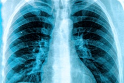
"Chest radiographs in particular have biological 'fingerprints' that can be used to identify a patient's images from a large number of similar images. These are the lung apex, the superior mediastinum, the cardiac shadow, thoracic fields, and the right lower lung," Morishita explained in a poster presentation at the 2008 American Academy of Physicists in Medicine's (AAPM) meeting in Houston.
Morishita and colleagues at Kumamoto University in Kumamoto, Japan; Iwate Prefectural Central Hospital in Morioka, Japan; and the University of Chicago in Illinois developed an automated method to search and compare a misidentified chest radiograph with a database of correctly identified chest radiographs in a PACS.
The team compared 200 chest radiographs, evenly divided between female and male patients, with an existing database of 26,210 chest radiographs. The PACS database included 20,267 exams of female patients and 15,943 exams of male patients, all of whom participated in a lung cancer screening program in Iwate, Japan.
For each chest radiograph, the five biological fingerprints (lung apex, superior mediastinum, cardiac shadow, thoracic fields, and right lower lung) were extracted. They were then used as templates to determine a correlation value between the misfiled image and each of the images in the database. The summation of the correlation values for the five biological fingerprints was calculated as a correlation index.
The automated search engine calculates the similarity of the correlation index values. If biological fingerprints in two images are identical, the correlation value is 1.0.
Morishita noted that "157 of the 200 unidentified images were correctly identified with a patient's chest images in the database."
"The search-and-compare algorithm was correct with 78.5% of the images. Moreover, it was possible to identify an additional 21 images, or 10.5%, when the top 10 patients whose radiographs were identified as most similar were compared in this subset," Morishita stated. "The ability for a computer to accurately identify images among a limited number of patients shows great utility by itself."
Some radiographs stumped the image-matching algorithm. It failed to provide high correlation values for 22 of the chest radiographs, even when they were compared with a dataset of images of the matching patients. Nonetheless, Morishita and colleagues believe that the study demonstrates the potential of utilizing biological fingerprints in diagnostic images to identify "orphaned" images in a PACS.
By Cynthia Keen
AuntMinnie.com staff writer
August 20, 2008
Related Reading
PACS data-mining tool analyzes CR retake rates, August 5, 2008
Copyright © 2008 AuntMinnie.com


