When viewing comparison images, the magnification of one image compared to another can affect radiologists' ability to perceive lesion growth, according to researchers from the University of Alabama at Birmingham (UAB).
"PACS vendors should offer an option to automatically display images at equal magnification," said Dr. Franklin Tessler, chief of body imaging at UAB. He presented the team's findings during a scientific session at the 2007 RSNA meeting in Chicago.
Clinicians make critical decisions every day about treatment based on changes in lesion size depicted on cross-sectional imaging. While it's long been possible to make direct linear measurements of lesions on the CT console, using that technique to track lesions is often impractical in the clinical setting, Tessler said.
"For example, if you're trying to follow a lesion over time, the technologist would have to know which lesion was being followed by reviewing prior studies before doing any measurement," he said. "And that really is difficult to do."
In the film era, radiologists could take advantage of the scale printed on every image or utilize a pair of calipers to compare lesion size growth. Most of the time, though, simpler methods such as with pen and paper were used, Tessler said. Thankfully, the advent of PACS has enabled radiologists to make direct measurements themselves on workstations, he said.
"But do we do this all of the time?" he said. "What happens if there are multiple lesions? Well, I dare say that most radiologists, when there are more than just a few lesions on a particular case, will measure some of them, particularly if they've been measured before. But if there are many lesions, we rely on a gestalt impression of growth, or decrease in size of the lesion without actually performing real measurements."
Much of the time, radiologists using PACS choose how to magnify images based on their own preferences. While some radiologists are comfortable performing size comparisons with images at different display sizes, many try to make the images as close as possible in size by either manually zooming or dezooming one of the images to get them roughly equal, Tessler said.
"Unfortunately, doing so is really subjective and often there is still some difference in magnification size from one image to the other," he said.
With that in mind, the UAB study team sought to determine the effect of the magnification of one image on the comparison of lesions over time. Prior research in the literature looking at simulated brain lesions on MRI had found that all three reviewers could detect differences if the diameter of one lesion was at least 15% greater than the other (American Journal of Roentgenology, January 2003, Vol. 180:1, pp. 65-69). In another study, researchers found that CT images magnified at one and two times did not improve performance and increased variability (Academic Radiology, April 2006, Vol. 13:4, pp. 407-408).
From the institution's PACS, the UAB study team selected 90 pairs of CT images depicting well-defined, hypodense liver lesions greater than 1 cm at two different points in time. For inclusion in the study, only one lesion could be on the image.
Each image pair was shown blindly to each of three abdominal radiologists with at least six years of experience in abdominal radiology. The radiologists each reviewed the image pair three times in one session, with both images at the identical size scale, with one image magnified by a factor of 1.69 (1.3 in either direction), and with the other image magnified by the same factor. The pairs were presented in a random order.
The radiologists were asked to indicate whether either lesion was larger or if they saw no difference in lesion size. Their responses were recorded by software, and the data were exported for statistical analysis.
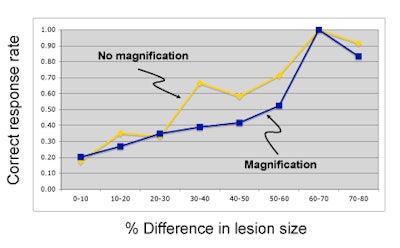
"The first main conclusion or finding was that obvious differences (in lesion size) were obvious," Tessler said. "But we also found that magnification tended to reduce performance."
 |
| Image courtesy of Dr. Franklin Tessler. |
At the midrange of differences in lesion size, there was a trend toward better performance with no magnification of either image, he said. Overall, however, that difference was not statistically significant.
Tessler acknowledged limitations of the study, including variable lesion definition, difficulty measuring the "true size" of the lesion, an uncertain threshold for real growth, the choice of magnification factor, and an unequal lesion size difference distribution.
"Nonetheless, we feel that we have shown that there is a trend toward a magnification effect on size difference discrimination that warrants study," he said.
In future research, the UAB plans to conduct an additional study evaluating this topic, this time with simulated lesions. This will allow for overcoming some of this study's limitations, Tessler said.
By Erik L. Ridley
AuntMinnie.com staff writer
January 21, 2008
Related Reading
Clinicians still need access to older imaging studies, December 10, 2007
Radiologists' job stress linked with computer competence, survey finds, November 29, 2007
The 2007 PACSman Awards: Great expectations, November 29, 2007
Communicating abnormal findings to clinicians may not ensure follow-up, November 28, 2007
Consumer displays compare well with medical-grade displays, November 27, 2007
Copyright © 2008 AuntMinnie.com