Support for 3D sonography has been building for years, with advocates claiming that the technology offers improved efficiency and less operator dependence over conventional ultrasound.
But how does 3D ultrasound's ability to take data volumes and reconstruct any plane enhance 2D sonography's clinical utility? Dr. Beryl Benacerraf and colleagues at Brigham and Women's Hospital, Massachusetts General Hospital, and Harvard Medical School, all in Boston, investigated whether a 3D reconstructed coronal view of the uterus adds benefit to standard pelvic ultrasound in women with abnormal findings. The team presented its work in the American Journal of Roentgenology (March 2008, Vol. 190:3, pp. 626-629).
"It turns out that this reconstructed coronal view is the most helpful in imaging the uterus, and now it's accessible with 3D ultrasound," Benacerraf said.
Sixty-six consecutive women had 2D pelvic ultrasound followed by 3D coronal volume acquisition focused on the area of the endometrium taken longitudinally along the length of the uterus. All 11 sonographers used an automated 4- to 8-MHz transvaginal transducer on a Voluson Expert system (GE Healthcare, Chalfont St. Giles, U.K.) for both exams.
The study was first read using the information from the 2D exam. Then one of three radiologists created a 3D coronal view of the uterus using multiplanar reconstruction.
The radiologist then evaluated whether the 3D reconstructed view did the following:
- Helped make a diagnosis that wouldn't have been possible with conventional ultrasound
- Helped confirm a diagnosis the 2D scan had suggested
- Did not add any diagnostic information to the conventional ultrasound
Benacerraf and her team found that the 3D coronal view of the uterus and endometrium added key information to conventional sonography in 24% (16) of the 66 patients. In five of these 16, the 3D view contributed additional diagnostic information that the 2D exam hadn't provided; in the other 11 patients, the diagnosis suggested by the 2D scan was confirmed with the 3D coronal view data.
The researchers found that three types of patients benefited the most from the 3D coronal view: those with a history of infertility or a questionable uterine abnormality, those with an endometrium thickness of 5 mm or more, and those with uterine fibroids.
 |
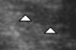
| A 31-year-old woman who presented with infertility. Above, transverse view through uterus shows no abnormality. Below, reconstructed coronal view of uterine cavity shows arcuate uterus. Benacerraf B, Shipp T, Bromely B, "Which Patients Benefit from a 3D Reconstructed Coronal View of the Uterus Added to Standard Routine 2D Pelvic Sonography?" (AJR 2008; 190:626-629). |
 |
For patients with an endometrium width of 5 mm or more, the 3D coronal view delivered either new data or data that confirmed a suspected diagnosis in 39% (11) of the 28 cases, compared to only four (12.5%) of 32 patients with an endometrium width less than 5 mm.
Thirty-four patients had fibroids that had been found with 2D ultrasound. In 24% (8) of these patients, the fibroids were seen more clearly with the 3D coronal view.
 |
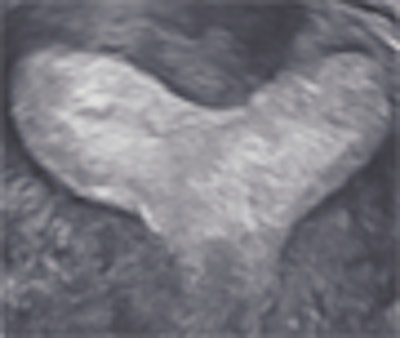
| A 40-year-old woman with a fibroid. Above, transverse view of uterus shows fibroid (calipers). Exact location of fibroid with respect to uterine cavity is unclear because posterior aspect of fibroid obscures side of endometrium. Below, coronal view shows that fibroid is not submucous, but is intramural without distorting shape of uterine cavity. Plane shown here was deemed by radiologist to be best plane to show closest relationship between fibroid and endometrium, showing that the fibroid did not extend into cavity but just abuts it. Benacerraf B, Shipp T, Bromely B, "Which Patients Benefit from a 3D Reconstructed Coronal View of the Uterus Added to Standard Routine 2D Pelvic Sonography?" (AJR 2008; 190:626-629). |
 |
The group acknowledged that the study's limitations included a lack of surgical confirmation for most of the cases, its small size, and the fact that the same sonographer who took the 3D volumes conducted the 2D scans. Yet Benacerraf and her group concluded that a 3D volume acquisition of the uterus offers valuable clinical data when used as an adjunct to 2D pelvic scans.
"The study shows that if there are suspicious findings on the conventional ultrasound, 3D can provide further information," Benacerraf said. "Ultrasound hasn't had the capability to take a volume of data and reconstruct it, like CT and MR have done for a long time. Now that it's possible to do this, it makes sonography more efficient and more complete."
By Kate Madden Yee
AuntMinnie.com staff writer
March 12, 2008
Related Reading
3D ultrasound offers efficiency improvements, March 29, 2007
3D brings opportunities to sonographers, August 4, 2006
2D ultrasound offers similar info to 3D/4D obstetric US, June 8, 2006
3D sonography of the endometrium adds value, May 5, 2006
Part II: The psychological impact of 3D ultrasound on pregnant women, December 30, 2003
Copyright © 2008 AuntMinnie.com