Exposure to dust produced from the collapse of the twin towers at the World Trade Center (WTC) on September 11, 2001, is subjecting the first responders to that disaster to an elevated risk of cardiovascular disease, according to results of a study involving dynamic contrast-enhanced MRI.
Researchers at Mount Sinai Medical Center have used the MR procedure along with peripheral arterial tonometry (PAT) to demonstrate that WTC emergency workers, who had relatively higher exposure to airborne pollution at the site, are also more likely to show early signs of atherosclerosis.
Extensive medical research has shown that exposure to hazardous substances pulverized during the collapse of the WTC buildings has contributed to high rates of cancer and respiratory disease among emergency personnel.
Dr. Mary Ann McLaughlin, an associate professor of cardiology at Mount Sinai, established in a study presented at the 2010 American Heart Association (AHA) annual meeting that the workers' cardiovascular health also warrants concern. She found diastolic dysfunctions among 60% of 1,200 first responders.
McLaughlin and colleagues expanded upon these findings at this year's AHA meeting in November. Their new study involved 31 ground zero workers recruited from the World Trade Center Medical Monitoring and Treatment Program. They were divided into two groups: The first group consisted of 19 subjects who arrived at ground zero on September 11, and the second group consisted of 12 workers who were first exposed to particulate matter at the site at least two days later. Their average age was 45.
PAT established a strong correlation between the severity of endothelial dysfunction in these subjects and their relative exposure to airborne pollution at the site. This loss of vascular elasticity is one of the first detectible symptoms of atherosclerotic disease, said the study's first author, Venkatesh Mani, PhD, in an interview with AuntMinnie.com.
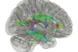
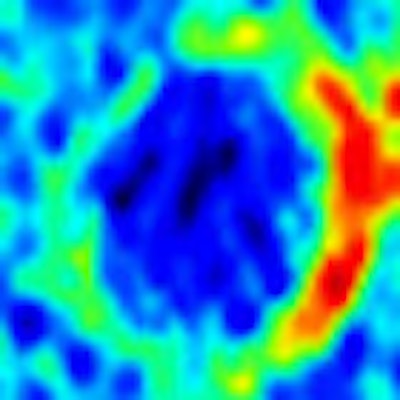
Dynamic contrast-enhanced MRI demonstrated that neovascularization, indicative of carotid wall inflammation and early arterial plaque development, was also more pronounced among workers exposed to more polluted air.
 |
| Sample dynamic contrast-enhanced MR images of left common carotid arteries reveal extensive neovascular development in a high-exposure patient (right) compared to a low-exposure patient. Images courtesy of Venkatesh Mani, PhD. |
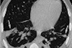
Black-blood MR angiography did not establish a significant difference in carotid plaque burden development between the two sets of workers.
 |
| No significant plaque burden appears in sample carotid images from patients with low (top) and high (bottom) particulate matter exposure. |
"Though this is a preliminary study, it gives us enough data to actually pursue this in the future in a larger population to verify our results," Mani said. "A lot more work is needed before this is applicable to everybody who was exposed."