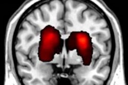
MRI with diffusion tensor tractography (DTT) could help physicians identify premature infants most at risk for cognitive, motor, and behavioral deficits.
The 3D MRI technique could also help them identify the type of deficit and begin early neuroprotective therapies, according to a study published online January 24 in PLOS One.
Researchers at Nationwide Children's Hospital in Columbus, OH, used DTT-MRI to detect abnormalities and subtle injuries in certain regions of the brain. The technique is thought to detect such structures and injuries better than earlier versions of the technology.
DTT also facilitated early and repeatable assessments of neuroprotective therapies to help clinicians determine whether treatments are effective in a matter of weeks, rather than the current lead time of two to five years.
Principal investigator Dr. Nehal Parikh, from the Center for Perinatal Research, and colleagues studied the development of 10 brain tracts in prematurely born infants. The tracts are associated with functions such as language, movement, and vision.
The group then compared the DTT scans to those of healthy, full-term newborns to confirm differences in the fibrous structure of the 10 tracts.
Although DTT-MRI is used regularly in adults, the tiny head size and lack of benchmark measurements in healthy infants meant that DTT for premature infants was previously uncharted territory. With the technique developed by Parikh and colleagues, DTT images can be reproducibly processed and reliably interpreted, according to a release from the hospital.