Researchers in Boston say that CT attenuation measurements can be used to distinguish untreated osteoblastic metastases from enostoses, which are benign bone lesions, according to a study in the August American Journal of Roentgenology.
The results mean that some biopsies and extra imaging studies could be avoided, the study team concluded (AJR, Vol. 207:2, pp. 362-368).
Imaging results can be confusing when an osteoblastic lesion is identified in the presence and absence of a primary malignancy, and the problem is exacerbated by the increase in incidentally detected lesions that comes with more CT scans being performed, said Dr. Connie Chang, from Massachusetts General Hospital's radiology department, in a statement.
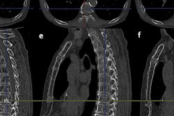
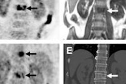
Aiming to distinguish between malignant and nonmalignant bone lesions, the research team examined 62 patients with 279 sclerotic bone lesions detected at CT, including 126 enostoses in 37 patients and 153 metastases in 25 patients.
While acknowledging the small sample size, the researchers concluded that a mean CT attenuation of 885 HU and a maximum attenuation of 1,050 HU offer reliable thresholds below which a metastatic lesion is likely to be the correct diagnosis.
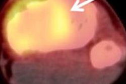
The detection of metastatic disease is important because it enables accurate diagnosis and determination of the course of care. Metastatic bone disease is the most common bone malignancy, affecting 400,000 people per year in the U.S.