Photodynamic therapy (PDT) is an effective and minimally invasive treatment for pancreatic cancer, and contrast-enhanced CT could potentially be used to predict PDT treatment response, according to a study published in Physics in Medicine and Biology.
The study team from Dartmouth aimed to reduce the imaging obstacles for safe and minimally invasive PDT treatment. Their results revealed a connection between the amount of blood in a tumor and its response to the light-activated treatment.
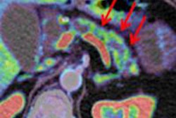
With photodynamic therapy, cancer cells are targeted using a drug called a photosensitizer. Cancer cells retain the photosensitizer longer than normal cells; when the drug is then exposed to light, it becomes active and leads to cell death. Because the response is chemically induced rather than heat induced, there is no damage to connective tissue.
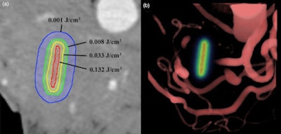
 A single axial slice (left) of the pancreas from the pretreatment CT scans is overlaid with computed contours of light fluence levels around the light-emitting fiber location. This was simulated using blood content information for tissue absorption from contrast CT. A volume rendering (right) of the blood vessels around the pancreas in the same patient is overlaid with the light dose map in the fiber location. Image courtesy of Dartmouth-Hitchcock Norris Cotton Cancer Center.
The researchers found that because blood absorbs light, if more blood is present, less light is available to trigger treatment; therefore, higher blood volumes reduced the effectiveness of PDT (Phys Med Biol, April 21, 2014, Vol. 59:8, pp. 1911-1921).
Consequently, the more contrast uptake the researchers saw on pretreatment images, the less the treatment had an effect, said co-author Brian Pogue, PhD, a professor at Dartmouth's Thayer School of Engineering and Geisel School of Medicine. This suggests that cancers with large blood volume and flow may be harder to treat with this therapy.
"This study implies that treatment response can be reliably predicted using contrast CT," Pogue said in a statement. "This would represent a major breakthrough in PDT for pancreas cancer that allows for easier, faster treatment tailored to an individual."