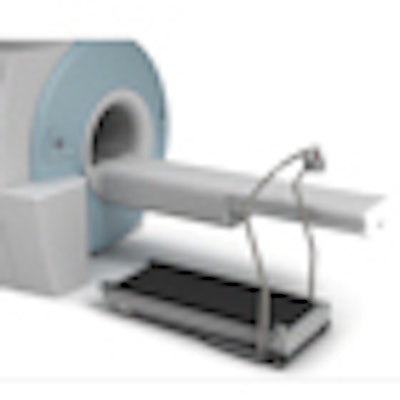
Researchers at Ohio State University (OSU) are achieving promising results with a specially designed treadmill for use in cardiac MR imaging to show exercise-induced cardiac wall-motion abnormalities.
The MRI-compatible treadmill can be safely located close to the scanner to minimize the distance between the treadmill and MRI gantry, enabling the scanning of patients immediately after exercise, when their heart rates are at peak stress. Orlando Simonetti, PhD, associate professor of internal medicine and radiology at OSU, spoke about the research at the International Society for Magnetic Resonance in Medicine (ISMRM) annual meeting this month.
From working at OSU's Ross Heart Hospital, Simonetti and his co-inventors found that cardiologists are interested in the key diagnostic and prognostic information that comes from a treadmill test, he said. However, MRI has traditionally been limited to pharmacological stress testing with the use of adenosine and dobutamine.
"A patient doesn't come into a hospital complaining they are short of breath when they infuse adenosine into their veins, or that they have chest pain when they inject dobutamine," Simonetti said. "It's when they are under exertion ... walking on the golf course or walking up the stairs that they have this chest pain. So, the goal of our test is to reproduce those symptoms and look at images that are potentially superior to SPECT or echocardiography."
Treadmill exercise
Simonetti noted that treadmill exercise is preferred to bicycle stress testing because the treadmill stress test provides a greater level of oxygen consumption. "On a treadmill, you can take a person to peak stress that is necessary to induce ischemia," he added.
The OSU team developed the MRI-compatible treadmill to sit next to the magnet and not interfere with its operation. Among the adaptations, Simonetti and colleagues replaced the drive system in the treadmill -- typically an electric motor -- with a hydraulic system through which energy is provided by a pump located in the MRI room.
"We are using an MRI-compatible, 12-lead electrocardiogram to monitor the patient, not while they are in the magnet, but while they are on the treadmill exercising," he added. "So it is really a conventional treadmill test in all aspects."
When a patient first arrives, he or she is given an MRI exam at rest. Cushions are placed on the gantry that mold to the patient's body. Immediately after the treadmill exercise, the patient is placed back into the mold and is automatically in position for clinicians to perform stress cine and stress perfusion MR imaging with no breath-holds.


"It is important that not only are the patients' respective heart rates at a level that indicates they are at maximum stress, but that the stress-induced defects have not disappeared," Simonetti said.
Clinical results
OSU researchers have tested the MRI treadmill on more than 100 patients. To gauge the efficacy of the technology, they evaluated 43 patients between the ages of 25 and 81 who underwent imaging procedures with both technetium-99m SPECT and gadolinium-enhanced cardiac MRI at stress and at rest.
The results, published July 2010 in the Journal of Cardiovascular Magnetic Resonance, found that stress cine cardiac MRI was completed in 68 ± 14 seconds following the end of exercise, while stress perfusion cardiac MRI was completed in 88 ± 8 seconds.
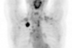
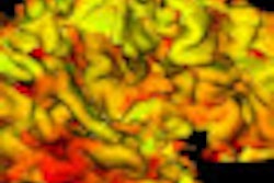
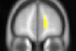
 First-pass MRI perfusion immediately following maximum treadmill exercise stress. Image indicates prominence of septal perfusion defect induced by exercise stress. Image courtesy of Orlando Simonetti, PhD.
Accuracy in patients who underwent coronary angiography was seven of eight (87%) patients for cardiac MR and five of eight (63%) patients for SPECT. Follow-up at six months indicated there were no cardiac events for 29 of 29 (100%) patients who had a negative cardiac MRI and for 33 of 34 (97%) patients with negative SPECT results.
The study, led by Dr. Subha Raman, concluded that exercise stress cardiac MR including wall motion and perfusion "is feasible in patients with suspected ischemic heart disease."
The authors added that larger clinical trials should be conducted to compare the efficacy of the stress imaging system with other stress imaging modalities.
Creating EXCMR
Simonetti and three other OSU co-inventors of the MRI treadmill have taken their technology toward the commercial arena by forming EXCMR. The company received a small business technology transfer grant from the U.S. National Institutes of Health in September 2009, just three months after hiring its first employee.
In August 2010, EXCMR was awarded a grant from the Ohio Third Frontier Medical Imaging Program, which provided $1.4 million and an additional $1.4 million in matching funds from collaborators.
The grant will fund a multicenter trial in which MRI-compatible treadmills will also be installed at Case Western Reserve University, University of Pittsburgh Medical Center, and the Christ Hospital in Cincinnati.
The trial will seek to scan approximately 450 patients with this technique in a head-to-head comparison with SPECT, using coronary angiography as the gold standard.