Diffusion-tensor MRI can have clinical value in measuring white-matter abnormalities and symptom severity in patients with obsessive compulsive disorder (OCD), according to a study published online April 7 in Radiology.
With the help of the MRI technique, researchers found that OCD is associated with microstructural abnormalities within the white matter of the brain that may indicate impaired integrity and increased connectivity. The lead author of the study is Fei Li, PhD, from the department of radiology at West China Hospital of Sichuan University.
Conventional T1-weighted MR images cannot garner enough information about white-matter integrity in the brain, even at high spatial resolution, the author noted. However, with diffusion-tensor MRI, the calculated fractional anisotropy contains information about directionality and coherence of neuronal fiber tracts, which affect OCD behavior.
The prospective study recruited 27 right-handed patients with OCD and 23 healthy right-handed patients between December 2007 and December 2008. All 50 subjects had similar demographics regarding age, gender, and education.
The researchers excluded potential participants who had, among other conditions, neurologic disorders, psychosurgery, current or past substance abuse or dependence, pregnancy, or any substantial physical illness, such as brain tumor, stroke, epilepsy, or hepatitis.
The final patient cohort consisted of 23 OCD patients and 23 healthy individuals. The OCD group included 16 male patients with a mean age of 24.4 years (range, 16-50) and seven female patients with a mean age of 33.4 years (range, 19-52). The healthy control group included 16 males with a mean age of 23.9 years (range,16-46) and seven females with a mean age of 33 years (range, 20-46).
Among the 23 OCD patients, 13 were receiving medication for OCD, while the remaining 10 were not receiving drugs at all. All 46 individuals were imaged on a 3-tesla MRI system (Excite, GE Healthcare) with the help of an eight-channel phased-array head coil.
Image analysis
Image preprocessing and statistical analysis were performed by three of the researchers, including Li.
Using a voxel-based analysis, the researchers evaluated the corpus callosum, which connects the right and left hemispheres of the brain and facilitates communication between the two regions, and the right superior frontal gyrus, which coordinates an individual's sensory system and self-awareness.
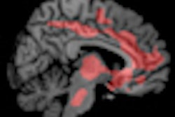
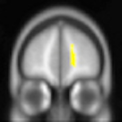
They found that OCD patients demonstrated significantly increased fractional anisotropy in the corpus callosum and in the white matter of the right superior frontal gyrus, compared with the healthy control subjects.
 |
| Parametric maps superimposed on 3D T1-weighted images show that OCD patients had significantly increased fractional anisotropy in the corpus callosum (above) and right superior frontal gyrus (below). Images courtesy of Radiology. |
 |
The mean fractional anisotropy for the corpus callosum in the OCD group was 0.499 ± 0.038, compared with 0.446 ± 0.039 in the control group. For the white matter of the right superior frontal gyrus, the mean fractional anisotropy in the OCD group was 0.411 ± 0.024, compared with 0.375 ± 0.025 among healthy subjects.
In the increased fractional anisotropy areas, axial diffusivity was greater in OCD patients compared with the control subjects, while radial diffusivity was not significantly different.
Li and colleagues noted the relatively small sample size as one limitation of the study, along with the fact that most patients had a history of medication prior to their MRI scans.
"Although the potential effects of medications on fractional anisotropy and axial and radial diffusivity are unclear," they wrote, "these issues need to be taken into account in future research."
A larger group of OCD patients who are not on medication should be recruited for long-term observation, the researchers added.
Despite the study's limitations, the findings suggest the potential clinical benefit of diffusion-tensor MRI to help monitor OCD progression and to indicate the need for clinical intervention, the group concluded.
"We found that OCD is associated with microstructural abnormalities of the white matter and a positive correlation between diffusion-tensor abnormalities and symptom severity, which may suggest that {diffusion-tensor] imaging might be of clinical value in measuring and following disability in OCD patients," they wrote.