Being identified as low risk on the Framingham cardiac risk assessment might convey a false sense of security if women are obese or have metabolic syndrome, say researchers from Massachusetts General Hospital in Boston.
A study of asymptomatic women found that individuals with those specific risk factors for heart disease have a high rate of cardiac abnormalities based on traditional risk assessments -- and that they should be screened with ultrasound. The research was presented at the American Society of Echocardiography (ASE) meeting in San Diego in June.
"We wanted to create a comprehensive program to reduce cardiac risk for women at high risk for heart disease," said Malissa Wood, MD, in an interview with AuntMinnie.com. Wood is a cardiologist and co-director of the Corrigan Women's Heart Health Program at the MGH Heart Center.
Both obesity and metabolic syndrome have been increasingly recognized as important risk factors for heart disease, according to the researchers. Ultrasound measures such as left atrial enlargement (LAE), left ventricular hypertrophy (LVH), diastolic dysfunction, and increased carotid intima-media thickness (IMT) have also been shown to portend worse cardiac outcomes.
The study -- led by Danya Dinwoodey, MD, with co-investigators including Lauren Gilstrap, MD, and Donna Slicis -- included 51 women ages 40 to 60. The women were obese and/or had metabolic syndrome, but they had no symptoms of heart disease and had normal heart function with echocardiography and ultrasound of the carotid arteries.
It is established that women with metabolic syndrome have a six- to sevenfold increased risk of heart disease. "In metabolic syndrome there are five features: If you have three, you make the diagnosis," Wood said. Those features include:
- Obesity
- Diabetes
- Glucose intolerance
- Elevated blood pressure
- Triglyceride to high-density lipoprotein (HDL) cholesterol ratio > 3
"We thought screening with ultrasound might be a good way to get a better risk assessment in women with these two risk factors," she said.
The Heart Awareness and Primary Prevention in Your Neighborhood (HAPPY) Heart Program recruited 51 asymptomatic low-income Caucasian and Hispanic women with two or more traditional cardiac risk factors and normal left ventricular ejection fraction to participate in the lifestyle intervention program.
Comprehensive echocardiography and ultrasound of the carotid arteries were performed at enrollment.
"We found, surprisingly, that a large percentage of the women ... had metabolic syndrome," Wood told AuntMinnie.com.
Of the 43 women who had complete ultrasound data, metabolic syndrome was represented in 32 (74%) women; the mean body mass index was 35.0 ± 8.2. Obesity was present in 29 (67%) of the women.
Nevertheless, all of the women had Framingham risk scores of less than 10%, putting them in the low-risk category.
"We found that Framingham doesn't perform very well in the women in our study," she said. In other words, "if you did a Framingham risk calculation they would come out as low-risk," but they actually had a high percentage of cardiac abnormalities.
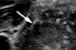
"In the women who met the diagnosis for metabolic syndrome, the majority had at least one abnormal ultrasound finding, either in the heart or in the carotid arteries," she said. "Chamber enlargement or thickness in the walls or carotid artery plaque -- if you do ultrasound in women you may find these other markers of disease risk."
Doing both tests -- i.e., echo and carotid ultrasound -- did reveal a number of cases that would have been missed if only one test had been performed, Wood said. The majority of patients had two abnormalities; typically, there was one abnormality in the heart and a second in the carotids, she said.
In addition, there was a clear correlation between the number of the five metabolic syndrome components and a greater number of abnormal ultrasound findings, including LAE, LVH, diastolic dysfunction, or increased carotid IMT or plaque.
Despite their being identified as low risk by traditional risk assessment tools such as the Framingham risk score, the study shows that women with obesity alone or the presence of metabolic syndrome have high rates of abnormal ultrasound findings, Wood said.
"You should consider using ultrasound as a tool in women who have these risk factors," she said. Screening women who have these two risk factors might allow for improved risk stratification and risk-reduction strategies, particularly in women identified as low risk.
For example, "currently, we do not recommend aspirin for those under age 65 to prevent heart disease, but if you see a women who has carotid plaque, you might recommend that she take a baby aspirin every day to reduce her risk," Wood said.
The center is continuing to collect data to confirm the results in more patients, Wood said.
By Eric Barnes
AuntMinnie.com staff writer
July 7, 2010
Related Reading
Cardiac model favors treadmill then CTA for low-risk patients, June 7, 2010
Trials aim to bring radiology ultrasound contrast to U.S., May 13, 2010
New CEUS technique images angiogenesis, March 1, 2010
Experience counts when reading contrast ultrasound liver images, January 19, 2010
Bracco revisits plans for U.S. launch of SonoVue, March 8, 2009
Copyright © 2010 AuntMinnie.com