Researchers from the University of Wisconsin aim to take radiation dose reduction to the next level in virtual colonoscopy.
Their study found that acquiring fewer images in CT scans would not render the virtual colonoscopy (also known as CT colonography) images nondiagnostic, thanks to the high intrinsic contrast between air and the colonic wall, and the clinical importance of the anatomy located at the interface of the colon wall and the lumen.
By undersampling the number of projections in the CT scans, they found they could theoretically reduce the dose at least fourfold without hampering image quality.
It's not that typical doses for a prone and supine virtual colonoscopy scan, running about 2 mSv or lower on average, are high to begin with, said lead investigator Matthew Christensen from the University of Wisconsin in Madison. "[CT colonography (CTC)] is really at an acceptable level, but if it can be done at a lower dose without compromising image quality, you would lower the risk even further," he said in a presentation earlier this month at the RSNA meeting in Chicago.
The study sought to demonstrate that adequate diagnosis in CT colonography can be achieved with fewer projections than are currently used, he said. Undersampling the number of projections without sacrificing imaging quality would reduce dose dramatically.
"The idea ... is that you have an image of a certain size, but only a small number of those pixels are actually important, and you can accurately reconstruct that image from a small number of samples randomly via a nonlinear acquisition procedure," he said. "Why this is interesting for CTC is that this is a unique type of CT exam in that ideally you would acquire a binary image. Air and colon are the only things you really care about."
In other words, only the interface between the lumen and the colonic wall is significant, which means there is a relatively small number of important pixels in the CT data, he said.
Christensen, along with colleagues Brian Nett; Jamey Weichert, Ph.D.; and Dr. Perry Pickhardt, loaded DICOM images from a typical CTC exam into Matlab software to be used as a digital phantom. They used the software's fanbeam function to forward-project the data using the geometry of a typical scanner.
"You take the CTC samples in a projection space, and you take enough projection samples to satisfy the Shannon-Nyquist theorem related to the number of detections that you have in the scanner," Christensen explained.
 |
| The Shannon-Nyquist theorem demonstrates that the scanner's analog signal can be reconstructed perfectly from the samples given a sufficiently high number of samples per the formula above, i.e., the number of projections (Nprojs) equals the number of detectors (Ndet) times pi over two. |
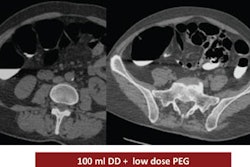
The sampled data became quite similar to the original CTC images acquired on a LightSpeed scanner (GE Healthcare, Chalfont St. Giles, U.K.), Christensen said.
For these images, the distance from the source to center was 541 mm, the number of detector elements was 813, and there were 1,200 views, the researchers wrote in an abstract.
The resulting sinogram was then decimated to form uniformly sampled projection datasets with undersampling factors of two, four, eight, and 16. The missing data were filled in using a linear interpolation technique and reconstructed using filtered back projections.
As a quantitative measure of the method, the root mean square error (RMSE) and signal-to-noise ratio of the reconstructed datasets were measured using the original as the reference.
To assess the data qualitatively, a gastrointestinal radiologist with significant expertise in CTC blindly reviewed the reconstructions as well as the original to compare diagnostic quality.
 |
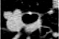
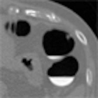
| There is little loss in image quality for undersampling factors of two and four. At eight, the image starts to degrade. All images courtesy of Matthew Christensen. |
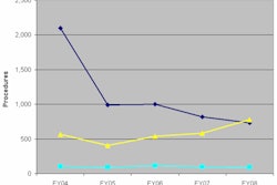
The results showed that the RMSE for the reconstructions -- decimated by a factor of two, four, eight, and 16 -- was 54.3, 124.9, 248.8, and 273, respectively. For the same factors, the signal-to-noise ratio was 27.3, 20, 14, and 13.2 decibels, respectively.
"There's little loss of image quality on undersampling factors [two and four], but once you get to a factor of eight the image starts to degrade," Christensen noted.
Qualitatively, radiologists couldn't tell the difference between the original images and an undersampling factor of two, but at undersampling factors of eight and 10, images were not considered diagnostic, Christensen said.
 |
| 2D image assessment by the radiologist could not distinguish between full reconstruction and those with undersampling factors of two and four for filtered back projection. Factors of eight and 10 resulted in a 2D filtered back-projection image that was not diagnostic quality. The RMSE rises with the undersampling factor. |
Filtered back projection wasn't the best reconstruction scheme, however. The researchers found substantially better image quality using an advanced reconstruction method they called compressed sensing.
Compressed sensing
"In compressed sensing, an image can be transformed into a compressed domain in wavelet [transform] or Fourier, which has sparse transform coefficients," Christensen said in his talk. "Each component in that domain is represented in all of these spatial [components] in the image domain, so by randomly sampling in that domain you might be able to capture the important information which allows you to accurately reconstruct it."
The method works best with random sampling, and unfortunately, fixed-geometry CT scanners don't allow for random sampling, Christensen said. MRI might actually perform better for this application.
However, because the critical information in the CT data lies at the boundary of air and tissue, relatively high spatial frequency components, the hope is that the data in the projection space will be sufficient to reconstruct the image, he said. In the group's experiment, details of which were not released to AuntMinnie.com, compressed sensing significantly outperformed filtered back projection as a reconstruction method.
Reduced sampling feasible
The researchers concluded that diagnostic quality appears to be achievable even with a fourfold reduction in the number of projections with interpolation (from 1,200 to 300), Christensen said. Further improvements in image quality are expected with the use of more sophisticated image reconstruction techniques such as compressed sensing.
The results support the notion that undersampling of virtual colonoscopy data can reduce dose substantially without compromising clinical diagnosis, he said.
But you won't be able to try this at home. In fact, you couldn't recreate the researchers' simulated dose-reduction technique on a real scanner if you wanted to -- at least not yet. Reduced sampling on CT would require hardware capable of switching the x-ray beam on and off rapidly.
Even that development may not be far off, though. Manufacturers are working on rapid switching capabilities for CT scanners, Christensen said.
By Eric Barnes
AuntMinie.com staff writer
December 19, 2008
Related Reading
Smaller teens sometimes get adult-sized CT dose, study finds, December 11, 2008
Studies address need to reduce CT dose in emergency settings, October 17, 2008
VC radiation dose holds steady overall despite dose modulation, June 2, 2008
Z-axis modulation cuts VC dose, keeps image quality high, April 12, 2005
Even low-dose VC yields extracolonic findings, September 21, 2004
Copyright © 2008 AuntMinnie.com