In musculoskeletal imaging, the modality of choice isn’t always readily apparent. To help, Dr. Levon Nazarian, a professor and vice chairman of education at Thomas Jefferson University (TJU) Hospital in Philadelphia, led an analysis of the existing literature and presented the findings at the 2006 Leading Edge in Diagnostic Ultrasound conference in Atlantic City.
Shoulder
Nazarian first discussed a TJU meta-analysis examining the accuracy of ultrasound and MRI in rotator cuff tears. The TJU researchers, led by resident Dr. Joseph de Jesus, retrieved 140 relevant articles, 28 of which met the meta-analysis inclusion criteria, which included the performance of imaging by radiologists.
Of the 28 studies, 20 examined only one modality, while eight were multimodality comparisons. The researchers found that ultrasound produced a summary sensitivity of 94.3% and a specificity of 95.3% for full-thickness tears, compared with 91.2% and 94.2% for MRI. MR arthrography offered sensitivity of 100% and a specificity of 92.9%.
"For full-thickness tears, there was no statistical significance in ultrasound, MRI, and MR arthrography," he said.
For partial-thickness tears, ultrasound produced summary sensitivity of 79.1% and specificity of 94.6%, while MRI turned in sensitivity of 63.1% and a specificity of 93.7%. The difference in sensitivity was statistically significant.
For all tears, ultrasound yielded summary sensitivity of 91% and a specificity of 87%. MRI produced sensitivity of 79.6% and a specificity of 90.6%. MR arthrography turned in sensitivity of 83.8% and specificity of 94.6%.
That study found that ultrasound is more sensitive than, and equally specific to, MRI in diagnosing a full- or partial-thickness rotator cuff tear, Nazarian said.
"We propose ultrasound as the initial imaging modality for rotator cuff tears," he said.
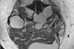
MRI should be the test of choice for labral tears, ligamentous injuries, fractures, and bone tumors, according to Nazarian.
Dr. Jon Jacobson, a musculoskeletal radiologist at the University of Michigan in Ann Arbor has suggested a shoulder pain algorithm in which patients less than 40 years old initially receive an MR or MR arthrogram, and patients older than 40 begin with ultrasound.
"You always have MRI as a backup," Nazarian said. Patients with dynamic impingement should also start with ultrasound, he added.
Elbow
For lateral epicondylitis, a prospectively blinded comparison found ultrasound’s sensitivity to range between 64% and 82% and specificity ranging between 67% and 100%. MRI yielded a sensitivity range of 90% to 100%, and a specificity of 83% to 100% (Journal of Clinical Ultrasound, May 2002, Vol.30:4, pp. 193-202).
In another elbow study involving eight baseball pitchers with medial elbow pain and a clinical suspicion of ulnar collateral ligament (UCL) injury, researchers found that grayscale ultrasound correlated with MR in 6 of 8 cases and with surgery in 3 of 8 cases (Skeletal Radiology, July 2004, Vol. 33:7, pp. 386-391).
Four UCL ruptures were diagnosed, which correlated with MRI in 4 of 4 cases and surgery in 2 of 2 cases. Two avulsions from the medial epicondyle were found, which were surgically correlated in 1 of 1 case. One sprain and one small partial tear were diagnosed, and those findings were correlated with MR in both cases.
"It appears from this study that MRI and ultrasound are about as accurate," Nazarian said.
In another UCL case series involving two symptomatic baseball pitchers, MR suggested a complete or partial UCL tear. Dynamic ultrasound was performed, and in one case the UCL was determined to be intact. The second case was confirmed as a complete tear (Skeletal Radiology, November 2002, Vol. 31:11, pp. 671-676).
"To summarize in the elbow, I would recommend ultrasound for ligamentous injuries because of the fact that you can do dynamic studies," he said. "Again, you do want to reserve MRI to look for associated pathologies too."
Ultrasound is recommended for nerve entrapments as well as tendon pathology, although MRI may be more sensitive due to adjacent bony abnormalities, he said. MRI is recommended for osteochondral injuries and loose bodies.
Wrist/hand
In one carpal tunnel study, ultrasound showed increased median nerve cross-sectional area in 16 of 20 cases, increased median nerve flattening ratio in 13 of 20 cases, and displacement of the flexor retinaculum in 9/20 cases. That study concluded that MR may show mild compression not seen with ultrasound, and that median nerve edema at MR aids in diagnosis (American Journal of Roentgenology, October 1992, Vol. 159:4, pp. 793-798)
Nonetheless, the researchers also concluded that ultrasound was an effective, low-cost, and widely available first study, Nazarian said.
In a 2004 study, researchers employing the 10 mm3 proximal to inlet and 12 mm2 to outlet criteria found ultrasound yielded a sensitivity of 86% and a specificity of 74% compared to electromyography. Interobserver variability was 0.87 to the tunnel inlet, 0.71 at the tunnel inlet, and 0.90 at the tunnel outlet. Another study found strong results for ultrasound compared with nerve conduction studies (Radiology, July 2004, Vol. 232:1, pp. 93-99; Rheumatology, July 2004, Vol. 43:7, pp. 887-895).
In an analysis of thumb UCL tears in 17 patients, a 1995 study found US to be 88% sensitive compared with surgery, while MRI was 100% sensitive even though a low-field magnet was used (Radiology, January 1995, Vol. 194:1 pp. 65-71).
For displaced tears, ultrasound was 83% specific, while MRI was 100% specific. In non-displaced tears, ultrasound was 91% specific and MRI was 100% specific.
"This may be a case where ultrasound could be done first, and if it’s equivocal, then go to MRI," Nazarian said.
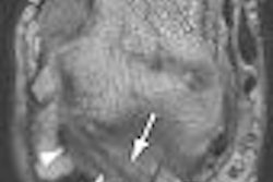
In a study examining scaphoid fractures, ultrasound produced 87% accuracy, 78% sensitivity, 100% specificity, 100% positive predictive value, and 75% negative predictive value when compared to the 1-tesla MRI gold standard. The modality was more accurate and more sensitive than x-ray (Radiology, July 2001, Vol. 220:1 pp. 231-235).
"If you’re thinking of doing an x-ray, you should also think of doing an ultrasound if you have experience with these fractures, because there is evidence that (ultrasound) is more sensitive than radiography," he said.
In a 2004 study involving triangular fibrocartilage in the wrist, ultrasound had an 88% correlation with arthroscopy and an 85% correlation with MRI. One tear was seen on ultrasound but not on MRI; there was no surgical correlation with that case, however (AJR, February 2004; Vol. 182:2 pp. 333-336).
For hand arthritis, MRI and ultrasound were found in a 1999 study to be more sensitive than radiography for inflammatory synovitis and bony erosions (Arthritis & Rheumatism, June 1999, Vol. 42:6 pp. 1232-1235).
"This is not even close," he said. "I cannot even tell you how many times that radiographs have been 100% negative, even in retrospect and read by bone radiologists who read bone films all the time every day, and you take (the patient) to ultrasound and they have huge erosions. It’s quite amazing. And of course, MRI is extremely sensitive for this as well."
In rheumatoid arthritis (RA), power Doppler ultrasound had a sensitivity of 89% and a specificity of 98% when compared to the gold standard of MR in detecting active RA (Arthritis & Rheumatism, September 2001, Vol. 44:9 pp. 2018-2023).
"We do use ultrasound as a screening (tool) for rheumatoid arthritis, for synovitis," he said.
In a comparison of ultrasound and MRI for occult dorsal ganglions, a 1994 study concluded that ultrasound should be the initial exam in suspected cases (Radiology, October 1994, Vol. 193:1, pp. 259-262).
For the wrist/hand, ultrasound is recommended for tendons, carpal tunnel, ganglion cysts, and synovitis (MR in equivocal cases), Nazarian said. Ligaments remain a gray area, however.
"I think it’s unclear which one we should do in the wrist," he said. "Until we’re a little clearer on what should be done first, I still feel more comfortable with (MRI in) triangular fibrocartilage and ligaments in the wrist, but as more data come in, we may find that ultrasound may be OK for that too."
By Erik L. RidleyAuntMinnie.com staff writer
September 7, 2006
Part II will examine US versus MRI in hip, knee, and ankle/foot musculoskeletal imaging.
Related Reading
From tears to TKA: The ins and outs of knee MRI, September 5, 2006
Color Doppler US shines for carpal tunnel syndrome, May 18, 2006
US with microconvex probes boost meniscal tear diagnosis, May 10, 2006
Focused exams suitable for some musculoskeletal US studies, March 26, 2006
US-guided needle tenotomy benefits tennis elbow, January 24, 2006
Copyright © 2006 AuntMinnie.com