The assessment of myocardial wall motion plays a key role in diagnostic and therapeutic decision-making. The sticking point is that wall motion is extremely difficult to assess quantitatively.
One common method, semiquantitative grading of regional wall motion with MRI, produces subjective results in the diagnosis of ischemia or myocardial viability, and reproducibility is limited, wrote the authors of a new study in Radiology.
Moreover, ultrasound methods such as tissue Doppler echocardiography, strain imaging, and strain-rate imaging with echocardiography are highly dependent on the transducer angle, in addition to other inherent limitations. Tagging methods obliterate lines or grids of signal as they move during the cardiac cycle.
But physicians from Oxford University in the U.K. believe they've found a comprehensive and reproducible method of assessing regional wall motion in tissue with phase mapping (TPM) of MRI data, a technique based on phase velocity.
"Phase-velocity-based methods allow direct observation of the velocity of the tissue by using the phase of the signal ... and allow determination of the displacement with integration over time," wrote Dr. Steffen Petersen, Ph.D., and colleagues (Radiology, March 2006, Vol. 238:3, pp. 816-826).
"Traditionally, TPM methods have been used to measure the displacement during short intervals and yield measurements of instantaneous velocity; the interval can be extended with use of the displacement encoding with stimulated echoes (or DENSE) approach that allows measurement of displacement during most of the cardiac cycle," they wrote.
"Optical flow of cine MR images provides an alternative approach for defining cardiac velocity values and displacement, and with it, high-resolution cine MR images that are not encoded with additional displacement information are used," the researchers stated. "Computerized algorithms are used to generate motion fields that explain the evolution of the images from one frame to the next. Such an approach is an extension of the function that is performed naturally by the human brain."
The group hypothesized that TPM would enable reproducible quantitative measurements of motion parameters, and given the method's high spatial resolution, that it would show differences between endocardium and epicardium for radial, circumferential, and longitudinal velocity parameters.
The authors used cine phase-contrast velocity MRI to establish a database of normal 3D systolic and diastolic endocardial and epicardial velocity values for all myocardial segments.
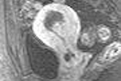
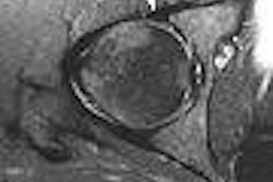
The group examined 96 healthy volunteers (57 men and 39 women; mean age 38 ± 12 years) with cardiac phase-contrast imaging on a 1.5-tesla scanner (Sonata, Siemens Medical Solutions, Erlangen, Germany). Steady-state free-precession images were acquired in horizontal and long-axis views (repetition time msec/echo time msec 3.0/1.5, flip angle 60º, section thickness 7 mm, gap 3 mm, temporal resolution 45 msec, 15 lines of k space per heartbeat).
Phase-contrast images were acquired with a black-blood segmented k-space gradient-echo sequence (6.2/4.5, flip angle 15º) with first-order compensation in all dimensions to minimize artifacts (pixel size 2.7 x 1.3 mm, 255 x 340-mm FOV, 96 x 256 matrix interpolated to 192 x 256, section thickness 8 mm).
Velocity encoding was performed by adding a bipolar gradient in the read, phase, and section directions to the otherwise identical sequence after each radiofrequency pulse, the group wrote. TR/TE was used to analyze 3D myocardial velocity on basal, midventricular and apical short-axis views.
Reproducibility was established by imaging 18 of the volunteers twice, and inter- and intraobserver variability were analyzed.
"Systolic and diastolic velocity curves were analyzed for peak velocity and time to peak velocity in the radial, circumferential, and longitudinal directions, as well as for torsion rate and longitudinal strain rate," they wrote.
The results showed that the TPM method provided a reproducible assessment of myocardial velocity with small intra- and interobserver variability.
"Systolic peak radial velocity was lowest at the apical level (p < 0.001); diastolic peak radial velocity was similar at all three myocardial levels (p = 0.73)," the team wrote. "As viewed from the apex, a relative counterclockwise rotation during systole was followed by a relative clockwise rotation of the apex against the base."
Diastolic and systolic peak longitudinal velocity values decreased from base to apex (p < 0.001). A gradient between endocardium and epicardium was observed from radial velocity values, with greater endocardial velocity values (p < 0.001).
"Contraction (positive radial velocity values) and expansion (relaxation, negative radial velocity values) patterns in the basal, midventricular, and apical levels were characterized by an increase in contraction velocity during early systole, followed by a slowing down of contraction velocity later in systole," the authors wrote. "The inverse pattern was observed in velocity. Similarly, an initial increase in radial expansion velocity values was followed by a decrease later during systole."
Of the two mature MRI-based methods of evaluating myocardial motion, TPM offers better spatial resolution, whereas tagging has better temporal resolution, "and may thus be superior in term of identification of peak deformation," the authors wrote. In any case, they explained, TPM data acquisition and postprocessing are quick and simple compared to complex and time-consuming tagging analysis. And TPM's temporal resolution could potentially be improved with the use of a navigator sequence, which would, however, increase imaging time.
Nevertheless, TPM is limited by lower temporal resolution compared to echocardiographically determined strain and strain rate and cardiovascular tagging. Moreover, echocardiographically triggered TPM "naturally misses about 10% to 20% of the cardiac cycle in late diastole," the group wrote, though they believe the shortcoming may improve with the use of high field strength magnets, improved gradient systems, and the implementation of parallel imaging.
The study results suggest that TPM is a reproducible comprehensive modality for assessment of regional wall motion. Intra and interobserver variability values were less than or equal to 5%, as calculated by dividing the standard deviation by the mean of the position of the section. And interobserver results in the study compared favorably to those obtained (approximately 15%) for echocardiographic strain and strain-rate imaging.
"Our results may potentially serve as reference data for which data from abnormal hearts can be compared, allowing TPM to be used as part of a multiparametric cardiovascular MR imaging approach in clinical practice," Petersen and colleagues wrote.
By Eric Barnes
AuntMinnie.com staff writer
April 3, 2006
Related Reading
Mental stress can induce ischemia in some CAD patients, March 17, 2006
PET, SPECT measures of LVEF have superior predictive value, March 13, 2006
Stress echo predicts risk in diabetics and nondiabetics, February 16, 2006
Rubidium-based PET may finally get its due, February 23, 2006
Viewpoint: 2D and Doppler echo spot diastolic heart failure, February 8, 2006
Copyright © 2006 AuntMinnie.com