Dual-time-point FDG-PET can aid in the differentiation of malignant from normal tissue in the breast, as well as in the detection of locoregional and distant metastasis from primary breast cancer, according to scientists from the Hospital of the University of Pennsylvania in Philadelphia. The technique involves scanning a patient at two distinct time intervals in order to quantify radiotracer uptake.
Dual-time-point FDG-PET can aid in the differentiation of malignant from normal tissue in the breast, as well as in the detection of locoregional and distant metastasis from primary breast cancer, according to scientists from the Hospital of the University of Pennsylvania in Philadelphia. The technique involves scanning a patient at two distinct time intervals in order to quantify radiotracer uptake.
"Biologic characteristics of the tumor such as histologic subtype (invasive-noninvasive)are considered important prognostic factors in patients with breast cancer and may therefore influence glucose metabolism as detected by 18F-FDG PET," wrote the authors in a study published in the Journal of Nuclear Medicine (September 2006, Vol. 47:9, pp. 1440-1446).
The researchers conducted a prospective study of 152 consecutive patients with newly diagnosed breast cancer ascertained via film-screen mammography, MRI, and biopsy. Patients who had biopsy performed underwent dual-time-point PET an average of 25 days after the procedure. Those patients who had hematoma, seroma, or inflammatory reaction to the biopsy were excluded from analysis, according to the scientists. They also noted that none of the study cohort had begun chemotherapy or radiation therapy prior to their PET scans.
The patients were imaged on an Allegro (Philips Medical Systems, Andover, MA) PET system, first with a neck-to-groin body scan, then a second set of chest images were obtained. The mean time interval between the injection of FDG and the first and second scans were 63 minutes and 104 minutes, respectively, and the patients did not leave the gantry between scans in an effort to minimize motion artifacts.
All patients underwent a surgical intervention after their PET scans, and the pathology result from the intervention was the gold standard against which the PET result was compared. A total of 32 patients were excluded from the study: 25 because biopsy had removed all tumor tissue and 7 because of 1- to 3-mm tumors, according to the researchers.
The images were reviewed by a nuclear medicine physician without comparison with other radiologic studies. Standardized uptake value (SUV) was measured from a region of interest (ROI) drawn around the site of a breast lesion on the PET images from the first time point to the second time point, and a percentage change was calculated. The patients were divided into two groups according to their pathology results: an invasive ductal, lobular, and mixed group, and those with carcinoma in situ. The invasive cancer group was further subdivided by lesion size to those greater than 10 mm and those with 4-10 mm lesions.
The visual results demonstrated 90.1% sensitivity for invasive cancers greater than 10 mm; 82.7% sensitivity for detecting invasive cancers 4–10 mm; and 76.9% sensitivity for detecting noninvasive breast cancer. Dual-time-point PET was true-positive for 99 lesions, indeterminate for 7, and false-negative for 17 lesions. The researchers noted that by visual assessment, most lesions became more intense on delayed scans.
 |
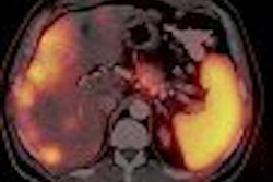
| Patient with invasive ductal carcinoma of right breast was examined with dual-time-point PET. (A) Coronal slices in top row were obtained at first time point. (B) Corresponding images in bottom row were acquired at second time point. Images in both sets clearly show primary lesion. However, intensity of uptake was substantially higher on delayed images. In addition, axillary lymph node metastasis was faintly visualized on first set but clearly demonstrated on second set. Measured SUVmax1 of lesion in first image set was 2.2 (thin black arrow), whereas that of second set was 2.6 (thick black arrow). Percentage increase in SUV of the lesion was 18.2%. SUVmax1 of metastatic right axillary lymph node was 1.0 in first set (open arrow in A) and increased to 1.1 in second set (open arrow in B). Surgical pathology confirmed 2.5-cm invasive ductal carcinoma with axillary metastasis. Copyright © by the Society of Nuclear Medicine. |
In the subgroup of invasive cancer patients, the team found an 8.6% increase in maximum SUV between the two scans for those tumors greater than 10 mm and a 6.5% increase in tumors between 4-10 mm. Invasive ductal tumors had an 8.1% increase, invasive lobular tumors showed a 10.5% increase, invasive mixed tumors had a 9.3% increase, and one medullary tumor demonstrated a 19.4% maximum SUV increase.
"Our results showed a strong correlation between dual-time-point changes and histologic subtypes in breast cancer," the authors wrote. "Another important result of our study was that, although the malignant tissues had positive dual-time changes in SUV, the normal breast tissue showed either no change or negative dual-time-point changes."
In a related study, researchers from the same institution examined the use of dual-time-point PET in detecting locoregional and distant metastatic lesions from primary breast cancer.
In this study, the group scanned 224 breast cancer patients with the dual-time-point technique and found 37 who were positive for locoregional or distant metastatic lesions, which were confirmed by histopathology or conventional radiological technique and clinical follow-up -- with the exception of lung lesions.
"Lung lesions which did not show an increase in SUV over time were proven to be benign inflammatory lesions," the authors wrote in a poster at the 2006 Society of Nuclear Medicine conference in San Diego.
They found that all lesions, other than those in the liver, demonstrated an increased uptake on delayed imaging, with the highest changes seen in the mediastinal and the internal mammary lymph nodes. In one patient who presented with three liver lesions, the SUV decreased between the first and second PET scan.
The scientists from both studies believe dual-time-point PET is the optimal method for SUV analysis for assessing suspected breast cancer. In addition, the increased uptake may be of help in differentiating benign abnormalities from malignant metastatic lesions, which could substantially improve the management of breast cancer patients, they wrote.
By Jonathan S. Batchelor
AuntMinnie.com staff writer
September 14, 2006
Related Reading
Preop FDG-PET spots axillary involvement in breast cancer, may obviate biopsy, August 21, 2006
PEM turns in solid results for detecting breast malignancies, November 10, 2005
PET mammography unit shows promise for DCIS, June 22, 2005
PEM development picks up pace, February 7, 2005
PET sharpens treatment plan for advanced breast cancer, August 16, 2004
Copyright © 2006 AuntMinnie.com