(Radiology Review) Multidetector-row CT (MDCT) of abdominal aortic aneurysms (AAA) treated with endovascular repair is best performed using 3-mm-thick images, according to a report in the December issue of the American Journal of Roentgenology.
Dr. Roberto Iezzi and colleagues from the University Gabriele d'Annunzio, Italy, compared the sensitivity, specificity, and positive predictive values of different slice thicknesses for detecting endoleaks (persistence of perigraft flow in the aneurysmal sac) at noninvasive MDCT.
"A successful EVAR (endovascular aneurysm repair) procedure depends on the complete sealing of the aneurysmal sac from blood flow to achieve general pressure relief and avoid aneurysm rupture, with shrinkage of the aneurysmal sac," they wrote.
Fifty consecutive patients with AAA treated with endovascular repair had contrast-enhanced four-slice MDCT scans using 1-mm collimation. Images were reconstructed using 1-mm, 3-mm, or 5-mm slice widths. As the study purpose was to evaluate arterial phase acquisition, accepted as the best phase for endoleak detection, delayed phase images were not analyzed.
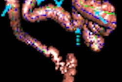
Endoleaks were classified using White's method: type I, incomplete sealing at the proximal or distal anchor site; type II, retrograde filling via aortic collateral arteries; type III, graft defect or a graft modules disconnection; type IV, graft wall porosity; or type V, increase in AAA size (endotension) without endoleak.
Triple-phase CT acquisition with and without contrast and 1-mm slice thickness arterial and delayed phase images were used as the reference standard for the detection and exclusion of EVAR endoleaks.
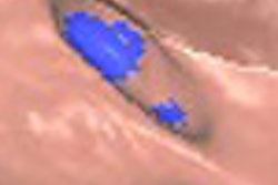
Eighteen (36%) of the 50 patients had an endoleak. One had a type I endoleak, 16 (88.9%) had a type II endoleak, and one had a type III endoleak. Two patients had type II low-flow leaks.
The authors found no statistically significant differences between the 1-mm and 3-mm image sets. However, the diagnostic accuracy for 1-mm and 3-mm image sets was significantly higher compared with the 5-mm image sets.
The department's surveillance program for EVAR patients includes clinical evaluation and triple-phase MDCT angiography at one-, six-, and 12-month intervals after surgery, then annual checks.
Although a small number patients had improved diagnosis with 1-mm image sets, this was not statistically significant. The 1-mm image sets have the disadvantage of increasing the number of images that must be interpreted and archived, the authors stated.
"On the basis of these results, we suggest that MDCT images obtained for follow-up after EVAR be reconstructed using a 3-mm slice thickness and 2-mm (reconstruction) increment," they concluded. "A second optional set of images should be reconstructed at a thinner thickness only in selected patients or in areas of concern, when the standard reconstruction protocol does not allow a definite diagnosis."
"MDCT Angiography in Abdominal Aortic Aneurysm Treated with Endovascular Repair: Diagnostic Impact of Slice Thickness on Detection of Endoleaks"
Iezzi, Roberto et al
Department of Clinical Science and Bioimaging, Section of Radiology, University "G. D'Annunzio," Chieti, via dei Vestini, Chieti, Italy
AJR 2007 (December); 189:1414-1420
By Radiology Review
January 14, 2008
Copyright © 2008 AuntMinnie.com