Cancer eventually recurs in more than a fifth of patients who undergo resection for renal cell carcinoma (RCC), according to radiologists from the University of Uslan in Seoul, South Korea. The most common recurrence site is the lungs, according to the researchers, who developed CT guidelines for detecting recurrent RCC based on a multiyear follow-up study of their own patients.
"In monitoring patients who have undergone surgery for RCC, radiologic examinations such as computed tomography (CT), radiography, and scintigraphy play a major role because physical examinations and laboratory studies are not sensitive for surveying tumor recurrence. Therefore, it is necessary to establish proper follow-up imaging strategies," wrote Dr. Eun Jin Chae, Dr. Jeong Kong Kim, and colleagues in the January issue of Radiology. "For this purpose, basic concepts or principles regarding the proper time and modality for follow-up examinations should be determined on the basis of knowledge about tumor recurrence patterns" (Radiology, January 2005, Vol. 234:1, pp. 189-196).
The retrospective study included 194 patients (162 men and 32 women, mean ages 54 and 56, respectively) who had undergone complete surgical resection of RCC with mean and median follow-up times of 45 and 79 months, respectively (range of seven to 92 months).
A rather complex exclusionary process narrowed the cohort to 194 patients for the retrospective study of hospital records. Excluded were those who had foci of metastasis at the time of surgery, while patients with regional lymph node metastases (N1 or N2) were left in. Also excluded were patients who missed follow-up sessions and those with incomplete tumor resection, more than one renal tumor, or any other malignant disease at the time of surgery, according to the authors.
Following administration of IV (iopamidol, Iopamiro 300, Bracco, Milan, Italy; or iopromide, Ultravist 300, Schering, Berlin) and oral (barium sulfate suspension, E-Z-CAT, E-Z-EM, Lake Success, NY) contrast material, CT imaging was performed using a Somatom Plus-S (Siemens Medical Solutions, Erlangen, Germany) or a 9800 Quick (GE Healthcare, Waukesha, WI) scanner, with a section thickness and reconstruction intervals of 5-8 mm. More lesions might have been found with the use of state-of-the-art scanners and thin-section imaging protocols, the authors noted.
Follow-up criteria included initial prospective CT exam within 12 months after surgery, and sequential CT exams every six to 12 months thereafter, as well as chest radiographs every three months during the first year after surgery and at least twice a year thereafter. Bone scintigraphy was also performed at six- to 12-month intervals, along with MRI or other imaging exams indicated by symptoms or clinical information.
Two radiologists evaluated the images by consensus, and recurrence was deemed to have occurred if a lesion grew in size based on imaging or was proved histologically, according to the First International Workshop on Renal Cell Carcinoma (Rochester, MN, 1997) held under the auspices of the World Health Organization, the authors wrote.
The results showed tumor recurrence in 41 (21%) patients, a mean of 17 months (range of three to 50 months) after surgery, and recurrence was seen within two years after surgery for 34/41 (83%) patients.
Tumor recurred within one year after surgery in 22 (54%) patients, within one to two years in 12 (29%) patients, within two to three years in one (2%) patient, and after three years in six (15%) patients, Chae and colleagues stated. "More than half of the tumor recurrences in the lung, bone, nephrectomy site, liver, and neck muscles were noted within two years after surgery. In particular, half or more of the tumor recurrences in lung and bone occurred within one year after surgery," they noted.
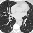
The recurrence sites included the lungs (n = 29, all detected by CT or radiography), bone (n = 13, seen on MRI in 10 patients and contrast-enhanced CT in seven), nephrectomy sites (n = 7, including two detected by ultrasound and seven by contrast-enhanced CT), brain (n = 8, including four detected by MRI and four by contrast-enhanced CT), liver (n = 5, including two detected by ultrasound, one by MR, and five by contrast-enhanced CT), mediastinal lymph nodes (n = 5), contralateral kidneys (n = 4), and the neck muscles (n = 2).
Patients with tumors 5 cm or larger saw recurrence more frequently than those with smaller lesions (p = 0.01), and recurrence was more common in patients with T3a or T3b tumors than in those with T1 tumors (p = 0.006 and p = 0.012, respectively). And those with nuclear grade 3 or 4 tumors saw a greater frequency of recurrence than those with grade 1 (p = 0.048 and p = 0.039, respectively) or grade 2 (p = 0.001 and p = 0.026, respectively) tumors, statistical analysis showed. On the other hand, "N stage and histologic subtype were not significantly related to the frequency of tumor recurrence," they wrote.
Follow-up early and often
Because surgery with or without systemic therapy has been shown to prevent localized tumor recurrence in selected patients with RCC, early detection of tumor recurrence may be useful in the goal of applying timely treatment, the authors wrote.
The recurrence patterns, in line with previous studies, showed that 83% of tumor recurrence (mean recurrence 17 months) occurred within two years of surgery, with considerable recurrence thereafter.
"Therefore, we suggest that follow-up imaging examinations should be intensively performed within two years after surgery, and that short-term surveillance after that period may be ineffective," they wrote. For example, 62% of bone metastases occurred within a year of surgery and 77% within two years, leading to a recommendation of regular bone follow-ups during the early postoperative period.
As the lungs are the most vulnerable site, chest CT should be performed in the early postoperative period, every six months during the first two years after surgery, then annually for two years in patients with a high risk of tumor recurrence, the authors concluded.
By Eric Barnes
AuntMinnie.com staff writer
January 20, 2005
Related Reading
US, MRI challenge CT for classifying renal cell carcinoma, June 17, 2003
Percutaneous radiofrequency ablation may be an option for solid renal masses, April 21, 2003
Radio-frequency ablation effective for many renal cell carcinomas, January 29, 2003
Copyright © 2005 AuntMinnie.com


