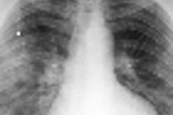
(Radiology Review) A comparison of 64-slice MDCT with intravascular ultrasound (IVUS) for coronary atherosclerotic plaques determined that 64-slice MDCT successfully identified disease, in particular for segments containing calcified plaques.
Radiologists at Beijing Anzhen Hospital in China studied 26 consecutive patients suspected of having coronary artery disease to determine whether MDCT improved individualized risk stratification.
Using both contrast-enhanced 64-slice MDCT (HP xw8200, Hewlett-Packard, Palo Alto, CA) and IVUS (20-MHz, electronic transducer, Volcano, Rancho Cordova, CA), Dr. Junyan Sun and colleagues analyzed images for presence, classification, and cross-sectional area percent stenosis of atherosclerotic plaques and compared the results for each modality (American Journal of Roentgenology, March 2008, Vol. 190:3, pp. 748-754).
Of 40 coronary arteries, 247 of 263 segments were imaged and analyzed by contrast-enhanced 64-slice MDCT using new plaque analysis software (SurePlaque, Toshiba Medical Systems, Tokyo) and IVUS, an accurate method for visualizing the wall of the coronary artery. With 16 segments excluded from the study because of poor CT image quality, 64-slice MDCT had a sensitivity of 97.4% and a specificity of 90.1%. Positive predictive value was 89.7% and negative predictive value was 97.5%.
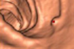
"Plaque analysis software using predetermined Hounsfield unit ranges for different components of plaque was able to distinguish between fibrous, fibrous-soft, and calcified plaques to a significant degree, but was less able to distinguish between soft and fibrous, and between soft and fibrous-soft plaque," they wrote.
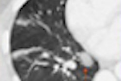
Imaging with 64-slice MDCT enabled high spatial and temporal resolution, and the semiautomatic plaque analysis software visualized and quantified both calcified and noncalcified plaque. Contrast-enhanced lumen, vessel wall, and even plaque morphology (soft, fibrous, or calcified) was color coded according to the CT Hounsfield unit.
The authors found that 64-slice MDCT is a valuable, noninvasive method for detecting, differentiating, and quantifying coronary atherosclerotic plaques and improves patients' risk stratification. However, they cautioned that "the reliable differentiation of the composition of individual noncalcified plaques is still limited."
"Identification and Quantification of Coronary Atherosclerotic Plaques: A Comparison of 64-MDCT and Intravascular Ultrasound"
Junyan Sun et al
Department of Radiology, Beijing Anzhen Hospital, Capital Medical University, An Ding Men Wai An Zhen Li, Chao Yang District, 100029, Beijing, China
AJR 2008 (March); 190:748-754
By Radiology Review
July 4, 2008
Copyright © 2008 AuntMinnie.com