Current clinical care of patients with spondyloarthritis is guided by criteria based on radiography findings, such as the modified New York criteria and Kellgren-Lawrence (KL) grading of radiographs of the sacroiliac joints. This is in spite of the fact that radiographs are difficult to interpret in cases of early disease, meaning some patients might go without therapy, according to the researchers.
Could CT be a better alternative? Researchers from the University of Alberta examined the records of 1,990 patients who received both radiographs and CT scans that included the sacroiliac joints over a five-year period. Readers reviewed the images and determined whether positive cases met the modified New York criteria and KL grading.
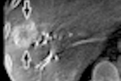
The researchers found nine patients with confirmed spondyloarthritis on both radiography and CT exams. However, only six of the patients met the clinical criteria on radiography studies, while eight met the guidelines for the CT scans. Agreement was poor for observations on x-ray, with kappa values ranging from -0.27 to +0.42, but it was better on CT, with an average kappa of 0.70 (range, 0.38 to 1.00).
Readers are much more likely to agree on features, diagnosis, and grading of sacroiliitis on CT than x-ray, and the implications of this for diagnosis of early spondyloarthritis call for the development of CT-based diagnostic criteria, the researchers concluded.