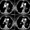
Pulmonary Edema:
View cases of pulmonary edema
Clinical:
Pulmonary edema is the abnormal accumulation of extravascular fluid within the lung parenchyma. Pulmonary edema may be secondary to high pulmonary capillary and venous hydrostatic pressure (cardiogenic pulmonary edema); or to increased capillary permeability with or without alveolar damage (non-cardiogenic pulmonary edema). Transudative edema (low protein content) is usually cardiogenic in nature, while exudative edema (high protein content [fluid-protein to plasma-protein ratio greater than 0.7]) is usually non-cardiogenic. Other signs indicative of non-cardiogenic pulmonary edema include a normal sized heart and vascular pedicle, lack of flow redistribution, no pleural effusion, and a patchy peripheral pattern of pulmonary edema
1. Cardiogenic Pulmonary Edema:
The most common cause of hydrostatic pulmonary edema is reduced left ventricular compliance and elevated end-diastolic pressures which are transmitted in a retrograde manner to the pulmonary circulation. Normal pulmonary capillary wedge pressure (PCWP) is less than 12 mm Hg. Redistribution of the pulmonary blood flow is seen when the wedge pressure increases to 16-19 mm Hg. Interstitial edema occurs at wedge pressures between 20-25 mm Hg, and alveolar edema is seen between 25-30 mm Hg. A reduction in PCWP is usually accompanied by significant clinical and radiographic improvement in these patients..
Hydrostatic pulmonary edema may occasionally be asymmetric, preferentially involving the dependent lung in a patient positioned in a decubitus manner. Pulmonary edema localized to the right upper lobe has been observed in patients with acute mitral valve incompetence and is most likely related to a jet effect of the regurgitant blood.
2. Non-cardiogenic Pulmonary Edema:
Some examples of non-cardiogenic pulmonary edema include:
- Allergic reactions
- ARDS
- Aspiration
- CNS injury: Permeability pulmonary edema without alveolar damage associated with CNS injury can occur in up to 50% of patients who have suffered a severe brain insult such as trauma, subarachnoid hemorrhage, stroke, or status epilepticus [9]. It usually presents acutely or within a few hours of the insult and resolves over several days. CNS pulmonary edema is a diagnosis of exclusion [9].
- Drowning/Near drowning: There is no significant difference in the radiologic features or clinical course following the aspiration of fresh or salt water. In both environments, chemical and organic contaminants in the aspirated water produce acute lung injury and resultant permeability edema. Initial chest radiographs often demonstrate scattered pulmonary opacities that progress to diffuse abnormalities over several days [8].
- Inhalational injury
- Hanta virus pulmonary syndrome
- Heroin overdose: Pulmonary edema develops in about 15% of cases of heroin overdose [9]. In this condition there is increased permeability edema without associated alveolar damage [9]. The edema resolves rapidly over one to two days [9].
- High altitude pulmonary edema: High altitude pulmonary edema results from endothelial leakage of fluid without alveolar damage [9]. It usually develops after 2 to 5 days at high altitude (over 3000 meters). The risk is increased with increasing altitude and rapidity of ascent. Young persons are also at greater risk. Patients generally improve rapidly when returned to a lower altitude and are given oxygen. In mild cases the edema is usually patchy, asymmetric, and spares the apices and bases. In more severe cases the edema is more diffuse. [5]
- Interleukin-2 or tumor necrosis factor administration: Causes increased capillary permeability without alveolar epithelial damage. Pulmonary edema occurs in about 75% of patients receiving IL-2 and in 15-20% of TNF treated patients [9]. Many patients are asymptomatic [9]. Radiographic findings are usually evident within 1 to 5 days of initiation of therapy [9]. The resulting edema is largely confined to the interstitium, small effusions are found in 40% of cases, and the findings clear rapidly [5,9].
- Re-expansion pulmonary edema
- Renal failure / Uremia: Pulmonary edema in patients with renal failure may be related to a combination of factors including expansion of the plasma volume (due to impaired salt and water excretion), a reduction in plasma oncotic pressure (due to protein loss in nephrotic syndrome), increased capillary permeability secondary to uremia, and left ventricular failure. These patients frequently demonstrate a widening of the vascular pedicle, cardiomegaly, and a classic central (bats wing) distribution of edema [2].
- Reperfusion edema after thromboendarterectomy: Reperfusion edema occurs in almost all patients following thromboendarterectomy for chronic pulmonary embolism. It is characterized by patchy bilateral perihilar alveolar opacities with maximum opacification generally seen by the second post-operative day. There is no correlation with preoperative pulmonary arterial pressures. [6]
- Severe hypoproteinemia/hypoalbuminemia (cirrhosis)
- Tocolytic therapy: Tocolytic agents are used to stop labor and include terbutaline, isoxsuprine, ritrodrine, and salbutamol [5]. Pulmonary edema develops in 4.4% of patients [5]. Increased risk is associated with underlying hypertension, diabetes, and coronary or valvular heart disease. The edema is usually bilateral and widespread. Pleural effusions may develop, but the edema usually clears rapidly after diuresis is begun [5].
- All-transretinoic acid (ATRA) syndrome: ATRA is used in the treatment of acute promyelocytic leukemia [10]. The agent can result in pulmonary edema in 22-25% of patients within 2 to 21 days (mean 7 days) of treatment [20]. Patients develop fever, fluid retention, hemorrhage, organ failure, thrombotic events, and usually a hyperleukocytosis with a rapid rise in leukocyte count [10]. Findings include enlarged heart, pleural and pericardial effusions, septal lines, and alveolar pulmonary infiltrates due to a capillary leak or pulmonary hemorrhage [10]. Treatment is withdrawl ATRA and steroids [10].
X-ray:
Cardiogenic (hydrostatic) pulmonary edema:
Plain film findings are well described and follow a logical progression from pulmonary vascular congestion (redistribution of pulmonary blood flow from the lower to upper lung zones), to a diffuse fine reticular pattern, to interstitial haziness, peribronchial thickening, and septal lines. Air space consolidation may eventually result with leakage of fluid into the alveolar air spaces. Initial radiographic findings may lag up to 12 hours behind acute changes in PCWP. A post-therapy lag is also observed and can range from hours to days following normalization of the PCWP as the edema fluid is resorbed.
Pulmonary vascular redistribution is most commonly associated with chronically elevated pulmonary venous pressures (ie: mitral stenosis or chronic left ventricular failure) and is less commonly observed in situations of acute elevations in pressure [5]. The typical patient with acute left ventricular failure will generally manifest mild lung edema and pleural effusions, but will not have dilated vessels or an enlarged heart [5]. Redistribution of flow can only be diagnosed on an erect film of the chest with a good inspiratory effort.
Interstitial pulmonary edema is demonstrated radiographically indistinctness of the vessels (early), the presence of septal or Kerley lines, and peribronchial cuffing or bronchial wall thickening [5]. Kerley B lines are short lines that are perpendicular and extend to the pleura. Kerley A lines are longer more centrally located lines that are often oriented more diagonally and cross normal vascular markings. Kerley C lines refer to randomly distributed superimposed linear opacities which produce a fine reticular pattern. Another finding of interstitial edema is thickening of the fissures which almost always precedes the development of septal lines [5]. As the amount of interstitial edema progresses, sufficient thickening of the interlobular septa may produce a hazy or ground glass appearance which is usually lower lobe or perihilar in distribution.
Fluid within the air spaces produces irregular, poorly defined areas of consolidation that are generally bilateral and tend to predominate in the central (bat's wing appearance due to sparing of the cortical areas) or lower lung zones. Pleural effusions are commonly associated with hydrostatic pulmonary edema and have traditionally been considered to be more common on the right (right 26%, left 16%, bilateral 59%). In chronic heart failure, cardiomegaly and pleural effusions can be present without visible vascular dilatation of lung edema [5].
Vascular pedicle: The vascular pedicle width (VPW) is increased in about two-thirds of patients with cardiac failure. The VPW is also commonly widened in patients with pulmonary edema secondary to renal failure or fluid overhydration. The corollary to this statement is also true- a less than normal VPW is strong evidence for diminished total blood volume such as dehydration or hemorrhage. The VPW is defined as the horizontal distance between the lateral aspect of the SVC shadow where it crosses the right main bronchus to a line drawn perpendicular to the lateral aspect of the left subclavian artery at its takeoff from the arch. The VPW is normally 38-58 mm (mean 48 mm) on an erect PA chest film. Inspiration and expiration have little effect upon the vascular pedicle width, however, rotation to the left will reduce VPW, and rotation to the right, will increase it. A film obtained at 40 inches (rather than 72 inches) will result in a geometric increase in the VPW of about 5% and the VPW will increase an average of 17% in a supine patient. However, only a small geometric increase is seen when changing from PA to AP positioning. [2,3,4] (Thanks to Dr. Craig Jonas, MD, for contributing articles to clarify the VPW)
In patients with COPD the findings of pulmonary edema may be less apparent. Due to the hyperaeration, changes in cardiac size may not be as demonstrated. Extensive parenchymal destruction in the upper lobes in these patients may mask vascular redistribution. Edema is usually most evident in areas in which the pulmonary parenchyma is preserved.
HRCT findings include pleural effusions, vascular redistribution, and cardiac enlargement. Other findings include diffuse or patchy areas of ground-glass opacification (which is seen early and may mimic an infectious process), interlobular septal thickening, centrilobular bronchovascular thickening, smooth perivascular interstitial thickening, increased vascular caliber, pleural effusion, or thickening of the fissures [1]. Pulmonary edema can also produce ill-defined centrilobular opacities which are typically associated with septal thickening. Enlarged mediastinal lymph nodes (up to 54% of patients) and hazy mediastinal fat (up to 35% of patients) have also been described in patients with congestive heart failure (CHF)- possibly related to edema within the nodes themselves [7]. The nodes are usually between 1-2 cm in size (short axis) and will decrease in size following resolution of the CHF [7]. Other etiologies for adenopathy should be entertained if the nodes are larger than 2 cm (even if CHF is present) and follow up scans should be obtained to ensure resolution of the adenopathy [7].
REFERENCES:
(2) Radiol Clin North Am 1991; 29(5): 943-963
(5) J Thorac Imag 1998; Ketai LH, Godwin JD. A new view of pulmonary edema and acute respiratory distress syndrome. 13: 147-171
(6) J Thorac Imaging 1998; Miller WT, et al. Reperfusion edema after thromboendartectomy: Radiologic patterns of disease. 13: 178-183
(7) AJR 1998; Slanetz PJ, et al. Mediastinal lymphadenopathy and hazy mediastinal fat: New CT findings of congestive heart failure. 171: 1307-1309
(8) AJR 1999; Marom EM, et al. The many faces of pulmonary aspiration. 172: 121-128
(9) Radiographics 1999; Gluecker T, et al. Clinical and radiologic features of pulmonary edema. 19: 1507-1531
(10) AJR 2002; Jung JI, et al. Radiologic features of all-trans-retinoic acid
syndrome. 178: 475-480


