Radiology 2003 Mar;226(3):749-55
Suspected pulmonary embolism: enhancement of pulmonary arteries at
deep-inspiration CT angiography--influence of patent foramen ovale and
atrial-septal defect.
Henk CB, Grampp S, Linnau KF, Thurnher MM, Czerny C, Herold CJ, Mostbeck GH.
PURPOSE: To investigate if abnormal early contrast enhancement of the aorta and
decreased attenuation of pulmonary arteries at deep-inspiration spiral computed
tomographic (CT) angiography might be caused by a patent foramen ovale (PFO).
MATERIALS AND METHODS: Two hundred forty-four spiral CT angiographic images of
the pulmonary arteries obtained during deep inspiration in patients suspected of
having pulmonary embolism (PE) were reviewed for evidence of abnormal early
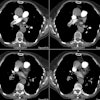
enhancement of the aorta. In 45 patients, enhancement of the ascending aorta was
equal to or more than that of the pulmonary arteries. Nonenhanced or contrast
material-enhanced echocardiography was performed in 39 of these cases. All CT
images with abnormal enhancement patterns were graded for contrast quality with
respect to sufficient enhancement of pulmonary arteries (four grades) at three
anatomic levels: right and left main and lobar and segmental branches. In
addition, all spiral CT angiographic images were evaluated concerning the
diagnosis of PE and the grouping of central (main pulmonary artery to proximal
lobar arteries) and peripheral (beyond proximal lobar branches) locations of
emboli. Mean attenuation values of ascending aortas and main pulmonary arteries
in group 1 (n = 244) were compared with those in groups 2 and 3 (n = 45) by
means of the two-tailed Student t test for unpaired data (P <.05). RESULTS:
Attenuation values for ascending aortas in group 1 were significantly lower than
those in groups 2 and 3 (P <.001). Attenuation values in main pulmonary
arteries were significantly higher in group 1 than in groups 2 and 3 (P
<.001). Echocardiographic images showed an intracardiac right-to-left shunt
in all 39 cases with abnormal contrast dynamics in the CT study (16% of the
whole study population). Three patients had an atrial-septal defect, and 36 had
a PFO. Images with a shunt had good (9%), intermediate (37%), fair (33%), and
poor (23%) contrast of the pulmonary arteries. Sufficient vessel contrast for
the diagnosis of PE could not be achieved in 27 of 45 patients with a shunt, but
severe central PE could be ruled out. PE could be diagnosed in 31% of the 244
images, 58% were negative, and 11% were indeterminate. CONCLUSION: A PFO may
frequently lead to insufficient attenuation of the pulmonary arteries, which
potentially limits the diagnosis of PE if the examination is performed during
deep inspiration.


