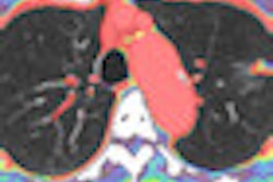
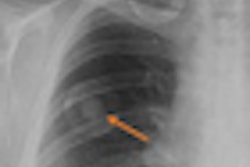
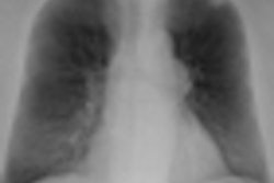
A prototype CT scanner with a flat-panel digital detector outperformed chest digital radiography (DR) in detecting pulmonary nodules, with more than twice the sensitivity of DR, according to Japanese researchers.
Flat-panel CT has intrigued researchers due to its high dynamic range, lower radiation dose than conventional CT, and compact design. Drawbacks include smaller fields-of-view and decreased temporal resolution, and the concept has yet to achieve routine clinical use.
Dr. Hideji Otani and colleagues at Shiga University of Medical Science developed the homegrown flat-panel CT prototype tested in their comparative study. The device consists of a 0.6-mm general purpose x-ray tube from Toshiba Medical Systems mounted on the ceiling, a vertically installed flat-panel x-ray detector from Canon designed for conebeam data acquisition, and a motorized rotating platform with a stool fixed almost in its center (Journal of Thoracic Imaging, February 7, 2011).
Twenty-six middle-aged patients, all but three harboring one or more pulmonary nodules, were enrolled. During flat-panel CT imaging, they were rotated while sitting on the stool, with the tube and detector fixed in a single position. Imaging parameters included 120-kVp tube voltage, 10-mA tube current, five-second exposure time, and total patient rotation of 720° including 180° for acceleration and 180° for deceleration. The frame rate was 200 frames per second, and the effective scanning area was 43 x 43 cm.
The same group was also examined for lung nodules with flat-panel digital radiography (DigitalDiagnost VS, Philips Healthcare) and again either with a 64-slice MDCT scanner (Somatom Sensation Cardiac, Siemens Healthcare) or 16-slice CT scanner (Aquilion 16, Toshiba).
Two radiologists identified nodules on MDCT by consensus to set a reference standard for comparing flat-panel CT and digital chest imaging. Four other chest specialists were blinded to the names and ages of patients when they read the flat-panel CT and anteroposterior digital chest images for pulmonary nodules.
A receiver operator characteristics (ROC) analysis established the superiority of flat-panel CT under most conditions (p = 0.02). The estimated mean Az value was 0.9818 ± 0.0083 on flat-panel CT images and 0.7610 ± 0.0908 on chest digital radiography.
The average sensitivity and positive predictive value (PPV) of flat-panel CT were 79.4% and 65.5%, respectively, compared with 33.8% sensitivity and 56.1% PPV for the digital chest radiographs. In all cases, the nodule-detection sensitivity of flat-panel CT was superior to chest radiography, Otani and colleagues wrote.
Flat-panel CT exposes patients to approximately three times more radiation than digital chest x-ray, but to considerably less radiation than chest CT, they noted.
Limitations of the flat-panel CT approach include the need for more time-consuming and careful interpretation of studies because of the less conspicuous presentation of lesions. Motion artifacts during patient rotation and insufficient radiation doses may result in strong streak artifacts relating to vertebrae or ribs. Also, patients must be able to maintain a steady sitting position.
Still, the findings on flat-panel CT are compelling enough to encourage adoption, according to Otani and colleagues.
"We expect this study to be the basis for the clinical use of [flat-panel CT]," they wrote.