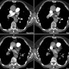
Segmental and subsegmental pulmonary arteries: evaluation with electron-beam versus spiral CT(1).
Schoepf UJ, Helmberger T, Holzknecht N, Kang DS, Bruening RD, Aydemir S, Becker CR, Muehling O, Knez A, Haberl R, Reiser MF
PURPOSE: To compare contrast agent-enhanced spiral and electron-beam computed tomography (CT) for the analysis of segmental and subsegmental pulmonary arteries. MATERIALS AND METHODS: CT angiography of the pulmonary arteries was performed in 56 patients to rule out pulmonary embolism. Electron-beam CT was performed in 28 patients. The other 28 patients underwent spiral CT with comparable scanning protocols. The depiction of segmental and subsegmental arteries was analyzed by three independent readers. The contrast enhancement in the main pulmonary artery was measured in each patient. RESULTS: Analysis was performed in 1,120 segmental and 2, 240 subsegmental arteries. One segmental (RA7, P =.010) and two subsegmental (LA7b, P =.029; RA6a+b, P =.038) arteries in paracardiac and basal segments of the lung were depicted significantly better with electron-beam CT. There was no statistically significant difference between electron-beam and spiral CT in the total number of analyzable peripheral arteries depicted. The mean contrast enhancement in the main pulmonary artery was 362 HU in electron-beam CT studies versus 248 HU in spiral CT studies. CONCLUSION: Detailed visualization of peripheral pulmonary arteries is well within the scope of advanced CT techniques. Electron-beam CT has minor advantages in analyzing paracardiac arteries, probably because of reduction of motion artifacts and higher contrast enhancement. Further studies are needed to establish whether electron-beam CT allows a more confident diagnosis of emboli in these vessels.


