Canadian researchers have found that FDG-PET/CT is more sensitive than MDCT in identifying recurrent and metastatic disease in patients with colorectal cancer and an elevated level of carcinoembryonic antigen (CEA), according to a study in the March issue of the American Journal of Roentgenology.
The study, performed at Princess Margaret Hospital in Toronto, also found that the two modalities have similar specificity in this clinical application (AJR, March 2010, Vol. 194:3, pp. 766-771).
According to the research team led by Dr. Ur Metser, previous research has not clearly defined the "benefit of FDG-PET/CT over MDCT in the assessment of tumor recurrence in patients with colorectal cancer and elevated CEA levels." CEA itself is used to monitor metastatic colorectal cancer and can be used to detect asymptomatic recurrences.
In their retrospective analysis, the researchers reviewed the histories of 158 patients who enlisted in a provincial PET registry trial over a 30-month period. All patients had a history of colorectal cancer, elevated or increasing CEA levels, and imaging scans that did not reveal an explanation for the elevated CEA.
Patient population
The researchers narrowed the study sample to 50 patients, who received a total of 55 FDG-PET/CT scans and underwent contrast-enhanced 64-slice MDCT of the chest, abdomen, and pelvis within 60 days of the FDG-PET/CT scans.
The sample group included 31 men and 19 women with a mean age of 61 years and a range of 28 to 89 years. All 50 patients received clinical follow-up for at least six months after the scans.
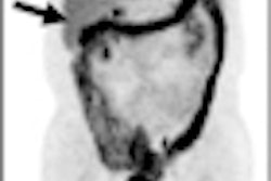
Whole-body FDG-PET was performed in 3D mode on a combination PET/CT scanner (Biograph, Siemens Healthcare, Malvern, PA). Patients were asked to fast for at least six hours before undergoing the scan, with data acquisition 60 to 90 minutes after an intravenous injection of approximately 5 MBq/kg body weight of FDG, up to 550 MBq.
Contrast-enhanced MDCT of the chest, abdomen, and pelvis was performed with a 64-slice MDCT scanner (Aquilion 64, Toshiba America Medical Systems, Tustin, CA).
Disease recurrence
In their analysis, researchers found recurrent or metastatic disease in 36 of 55 images (65.5%) when the patient also had an elevated CEA level. In addition, 54 of 61 tumor sites suspected as tumor recurrence with either imaging modality were found to be local recurrence or metastatic colorectal cancer.
In patients with elevated CEA levels, FDG-PET/CT registered a greater sensitivity than MDCT in identifying tumor recurrence, while the two modalities achieved similar specificity. FDG-PET/CT also scored greater sensitivity and specificity than MDCT in analyzing tumor locations.
|
FDG-PET/CT was able to correctly identify 17 tumors that MDCT did not detect. Both modalities prospectively missed one site of tumor recurrence.
Of the tumors identified by FDG-PET/CT and missed with MDCT, five were local recurrences in the presacral space, four were metastatic subcentimeter lymph nodes, three were peritoneal deposits, and three were recurrences at the periphery of radiofrequency-ablated metastatic lesions of the liver and in the abdominal wall. One was in the uterine cervix.
The authors noted several limitations of the study. They cited potential selection bias because the enrollment study did not choose consecutive patients with colorectal cancer and elevated tumor markers. Instead, the sample group was based on certain criteria and taken from the provincial PET registry trial.
"These results, therefore, may not be applicable to all patients with findings of colorectal cancer at imaging surveillance," the authors wrote. "Because of the selected patient sample and high pretest likelihood of the finding of tumor recurrence at PET/CT, it is possible that the sensitivity of PET/CT was overestimated."
Also, the FDG-PET/CT and MDCT scans were conducted within 60 days of each other, and it is "conceivable that this interval may have adversely affected the sensitivity of the earlier imaging study," the researchers noted. "However, the MDCT studies included did not necessarily precede PET/CT, and the group of studies with the shortest intervals between examinations was chosen to minimize bias in favor of one of the techniques."
By Wayne Forrest
AuntMinnie.com staff writer
February 22, 2010
Related Reading
PET/CT cost-effective for head, neck squamous cell carcinoma, February 17, 2010
PET/CT finds brain-disorder-related cancer other modalities miss, January 11, 2010
FDG-PET/CT helps manage treatment for Crohn's disease, November 16, 2009
Whole-body PET finds tumors missed by other tests, May 1, 2009
FDG-PET/CT shows early chemo results, April 16, 2009
Copyright © 2010 AuntMinnie.com