CHICAGO - If you're looking for microvessel obstruction in patients with acute myocardial infarction, it's worth the rush to perform cardiac MRI within 48 hours after the event -- and within two minutes of injecting the contrast. You could potentially help your patients live longer.
The microvessel obstructions indicated by the presence of "no-reflow" zones in those early post-MI scans carry potentially significant implications for patient care, researchers from Germany have concluded.
The motivation behind their work was studies conducted by Wu et al (Circulation, August 27, 2002, Vol. 106:9, pp. 1,083-1,089) showing that event-free survival was definitely determined by the infarct size and the presence of microvascular obstruction, according to Dr. Karl-Friedrich Kreitner from Johannes Gutenberg University in Mainz, Germany.
"In our prospective study, we tried to evaluate late-enhancement (LE) and no-reflow (microvascular obstruction or MO) zones in patients with acute myocardial infarction and successful recanalization with an acute stent (PTCA) with a one-year follow-up," he said in a presentation Thursday at the 2005 RSNA conference.
Group 1 (mean age 56) of the study cohort consisted of 40 patients who were examined with cardiac MR twice within the subacute phase -- once within 48 hours of MI symptom onset, and again within 10 days. There were 47 patients in group 2, only one of whom had cardiac MR within the first 10 days of symptom onset. This second group of patients underwent longer-term MRI follow-up at six and 12 months.
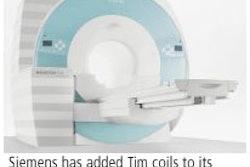
The exams were performed using a 1.5-tesla scanner (Magnetom Sonata, Siemens Medical Solutions, Erlangen, Germany) equipped with a six-channel phased-array coil. LE and MO were assessed using a 2D and 3D segmented TurboFlash sequence after the group determined optimal T1 in a short-axis orientation of the heart. All patients were injected with 0.2 mmols gd-DTPA kg/body weight for contrast enhancement, with images acquired after a scan delay of two minutes, Kreitner said.
"After image acquisition, we performed a planimetric quantification of areas of hyper- and hypoenhancement of the left ventricle on short-axis slices using commercially available software," he said.
For the group 1 patients, areas of late enhancement decreased significantly within the first 10 days of symptom onset (from 20.1% ± 11.6 to 16.7% ± 10.5, p < 0.05), Kreitner said. Areas of microvascular obstruction also decreased significantly during the same period (from 3.4% to 2.4%, p < 0.05).
For group 2 patients, late enhancement decreased by about 40% between subacute phase imaging and six months' follow-up, and in some cases LE continued to show a decrease as long as a year after MI.
Also in group 2, microvessel obstruction depicted as areas of MO decreased over the year (from 17.1% ± 11.42 to 6% ± 11.9, p < 0.05). The number of patients affected by MO was 29 in the subacute phase, 12 at six months, and just three at one year, he said.
In a woman examined 40 hours after AMI, "we could nicely see the microvascular obstruction," Kreitner said, displaying an image. In the same patient six months later, islands of low-signal-intensity tissue indicated a possible recovery of viable myocardial tissue in the region of the former infarct.
By far, the extent of MO was largest in images obtained two minutes after contrast administration. By eight minutes following contrast injection, the areas of visible MO were reduced dramatically, he said.
"The areas of late enhancement decrease from the acute to the subacute phase after acute MI, and this phenomenon can be demonstrated for some patients even after the time course of one year," Kreitner said. "For assessment of the prognostic parameters, we think it's essential that you try to examine those patients as early as possible, preferably within 48 hours after the event.
The underlying pathophysiologic phenomena are still being debated, he said, but his team believes that development from normal myocardial tissue to capillary leakage to membrane rupture is a progressive disease process. In between leakage and rupture, there may be a "membrane flaps" stage that is at least partially reversible, he said.
By Eric Barnes
AuntMinnie.com staff writer
December 1, 2005
Related Reading
Ventricular remodeling after AMI correlates with gelatinase activity, December 1, 2005
Five-year outcomes good after percutaneous coronary intervention for MI, November 29, 2005
Copyright © 2005 AuntMinnie.com