Patients who present with acute deep veinous thrombosis (DVT) of the lower extremities also have underlying anatomic problems, according to Korean interventional radiologists. The group, from Seoul National University Hospital, now routinely uses spiral CT venography to assess DVT patients.
One of the main goals of imaging DVT is for pre-treatment planning, said Dr. Jin Wook Chung in a presentation at the 2003 Society of Interventional Radiology meeting last month.
"In cathether-directed thrombolysis for DVT, the information about the extent of thrombosis, the type of thrombosis (acute versus chronic), the anatomic variation, and the underlying causes of thrombosis are necessary," Chung said.
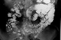
For the study, conducted over a four-year period, 30 patients with acute DVT were evaluated with spiral CT venography, performed with 3-mm beam collimation and a 2-mm reconstruction interval. Contrast media was injected via the antecubital veins and a spiral scan was initiated 5 minutes later. Precontrast images were obtained with 5-mm thickness and 10-mm intervals.
The images were analyzed using axial sections and 3-D reconstruction, including multiplanar reformatting and volume rendering. Chung and colleagues looked for the presence or absence of proximal obstructing lesions and their causes.
Of the 30 patients, 26 had left-sided DVT and four had right-sided DVT. Among those with left-sided DVT, 24 patients had stenosis or obstruction in the left common iliac vein. The causes of obstruction were as follows:
- 18 patients with compression by the right common iliac artery
- 2 patients with compression by the left common iliac artery
- 1 patient with an enlarged uterus
- 1 patient with combined causes.
In two patients, bony spurs along with iliac vein compression were the reason for the obstruction. This condition is commonly known as May-Thurner syndrome.
Among the four patients with right-sided DVT, three had stenosis or obstruction. One patient had right common iliac vein compression, between the right common iliac artery and spine, with a bony spur. One patient had segmental external iliac vein stenosis and one patient had femoral vein stenosis.
In all patients, the obstructing lesions demonstrated on spiral CT venography were judged to be true lesions during catheter-directed thrombolysis, Chung said.
"In conclusion, the majority of patients with spontaneous acute DVT in their lower extremity have underlying anatomic problems," he said. In answer to audience questions, Chung said the majority of patients were referred to the interventional radiology department because of severe leg edema. While the presence of bony spurs can alter the interventional method due to higher risk of artery erosion, Chung said his group routinely stents these patients who have complications.
In a commentary immediately following Chung’s talk, Dr. Charles Semba from Stanford University in Stanford, CA, praised the study, stating that it bolstered conventional wisdom that iliac vein compression will have an anatomic reason for obstruction.
"Your paper nicely demonstrates that May-Thurner syndrome is quite common. It really helps us in driving our therapy," Semba said, adding that the goal of therapy is to "debunk the lesion and look for underlying anatomic variations." These variations can help interventional radiologists decide if angioplasty and stenting is possible.
However, Chung cautioned that the patients in the study were, obviously, Asian, but that the etiology of DVT can differ by race.
By Shalmali PalAuntMinnie.com staff writer
April 9, 2003
Related Reading
New scintigraphy approach helps in DVT diagnosis, March 10, 2003
New techniques increase ultrasound’s value in vascular imaging, July 8, 2002
D-dimer assay plus baseline ultrasound accurate indicator of DVT, April 29, 2002
Combination of two bedside tests can exclude pulmonary embolism, July 28, 2001
Copyright © 2003 AuntMinnie.com