Professional baseball players in the U.S. face a long and demanding season. It starts with spring training in March, and, for a lucky few, ends with the World Series in October. All told, these athletes play nearly 200 games a season, with most games back-to-back, and about half away from home field -- and away from local imaging providers.
When a player is injured, the San Francisco Giants organization needs to know the extent of injury and the prognosis for rehabilitation -- as quickly as possible.
"For us, because there is a game every day, it’s not like football where they have seven days to get ready for the next game. Every day that we have to wait for a diagnostic decision, we lose that player for a day," said Stan Conte, the head trainer for the Giants. "Now, that may not sound like much, but we only have 25 players on a team and each game is critical -- so we need to know right away whether that player should be replaced. Not only do we have the health aspects of doing things right, but you also have the administrative properties of being able to make the right move for the team, both for the short term and long term."
Conte places great value on the role of radiology in assessing the health and rehabilitation of his injured players. He was one of the first trainers in professional baseball to make both digital and teleradiology an integral part of his sports medicine toolkit. Conte took some time out from the Giants spring training session last month in Tucson, AZ, to talk about the advances in imaging that have helped his team play ball.
Teleradiology scores a home run
"When we moved into (the new stadium) Pac Bell Park, I found out that our x-ray machine (Shimadzu Medical Systems North America, Torrance, CA) was hooked up to a CR machine (Eastman Kodak Health Imaging, Rochester, NY) that provided digital output of radiographic images," Conte said.
Conte decided to have the team’s healthcare provider, Catholic Healthcare West/Saint Francis Memorial Hospital of San Francisco, install a fiberoptic cable from the imaging equipment to the training room so that team management could look at the images immediately, he said.
Radiologic technologists at Saint Francis perform the x-ray exams on injured players at the ballpark.
"Our techs are using standard x-ray protocols for our work at Pac Bell Park. For example, for elbows we’re using a mAs of 10, a kVp of 70, and a 40-inch distance on a 10 x 12-inch receptor; when we’re imaging a forearm we use the same distance, mAs of 7, and a kVp of 75 on an 11 x 14-inch receptor," said Linda Womack, the hospital’s radiology department administrator.
Conte initially performed the fiberoptic hook-up to determine if there would be a benefit to viewing a digital image versus film. Once he saw the advantages of various image-enhancement algorithms available via the workstation software (Merge eFilm, Milwaukee), and the speed at which the images were available, his thoughts turned toward making the technology available in every city the team visits.
 |
|
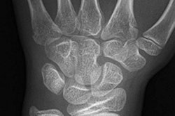
Hamate Fracture
History: A 20-year-old male outfielder who hits left-handed swung and missed the ball. He experienced immediate hypothenar eminence pain. Clinical examination showed tenderness over the muscle belly of the hypothenar eminence in the palm of the hand. No wrist pain or tenderness; demonstrated full range of motion in wrist and fingers. Grip strength was near normal the day after the injury. X-rays, including carpal-tunnel view, were inconclusive for pathology. MRI made the diagnosis of a fracture of the hook of the hamate. Significance: This injury almost always occurs in a hitter who swings and misses and has immediate ulnar palm pain. It is always the bottom hand with which the player holds the bat. This is the significance of this player being a left-handed batter. The bottom hand in the batting position is the right hand in a left-handed batter. The precise mechanism of injury is unclear, but theory has it that the hook of the hamate bangs mechanically against the knob of the bat. Treatment: The San Francisco Giants have found that most if not all such fractures deteriorate to non-union, becoming chronically symptomatic. Therefore, excision of the fractured hook is performed as soon as the diagnosis is made. Return to full competition is usually possible within 6 weeks, although players have returned as early as 3-4 weeks. Image and case study courtesy of the San Francisco Giants and Clinical Diagnostic Radiology, Phoenix. |
"X-rays are important, but our initial problem was with our MRIs because of the amount of data involved and the time it takes to transmit it," Conte said. "What we started out doing is, if somebody had an MRI, we would take the cuts that we wanted from the series and put it up on a lightbox, take digital pictures, and send them to our radiologist in the Bay Area. It would take forever, but at least our physicians back home would get some idea of the injury. Obviously, caveman technology, but it was a way to not have to wait for overnight delivery."
Dr. Charles Ho and associates at California Advanced Imaging perform the majority of MR imaging for the team, and they also read all other imaging exams. Conte and his crew have come to know and trust this Atherton-based radiology group over the years.
"I’ve found that radiologists are different, especially in how they read MRIs. It’s more of an art based on science in my mind than it is a ‘here’s a fracture, here’s not a fracture' scenario," Conte said.
From its primitive beginnings in teleradiology, the team has come to exclude the use of imaging centers in other cities that do not support Web-based image transfers.
"If, for instance, we’re in Pittsburgh, we use an MRI center that has the capability to electronically send the image files to Dr. Ho, who will then read the film and put it in his network," Conte said. "Then our orthopedist can access the images via the Internet and be able to look at them, because an orthopedist looks at an injury differently than a radiologist. So now we have, within an hour, two opinions that we value, and we’re able to determine exactly what we need to do next with the player. For us, it’s absolutely critical."
The Giants also have a set of MRI protocols for imaging that Ho has developed, and insist that these protocols be followed by any facilities that perform imaging procedures on the team’s players. According to Conte, arm injuries, particularly to wrists, shoulders, and elbows, are common among professional baseball players.
 |
|
Ulnar Collateral Ligament Tear of the Elbow
History: A 23-year-old male pitcher delivered a pitch and felt a very distinguishable "pop" in his elbow. He could not deliver another pitch and was immediately taken out of the game. Clinical examination showed obvious swelling of the medial aspect of the elbow with lack of flexion and extension. Tenderness was present along the medial elbow, but specifically just distal and posterior to the medial epicondyle. Valgus stress testing in 30° of flexion was very painful. MRI confirmed the diagnosis of complete tear of the ulnar collateral ligament. Significance: Although this can happen in any thrower, the injury occurs most often in pitchers. Interestingly, these acute injuries occur in game situations and not in practice, undoubtedly because of the velocity of the pitch. The throwing motion inherently places a valgus stress on the ulnar collateral ligament. Increasing the velocity increases the valgus stress, and is most likely why it occurs in games. Treatment: Acute ruptures of the ulnar collateral ligament are treated with surgical reconstruction, usually utilizing the palmaris longus, although other tendons can be used. Primary repairs have been tried, and some have been successful, but the so-called "Tommy John" surgery (a procedure wherein a tendon is taken from the non-pitching arm to replace the torn ligament) is the gold standard for this injury. Return to competition usually takes 12-16 months. Image and case study courtesy of the San Francisco Giants. |
MRI as MVP
At California Advanced Imaging, all scans are performed on a Symphony 1.5-tesla MR scanner (Siemens Medical Solutions Malvern, PA). Susan Bybee, chief technologist for California Advanced Imaging, shared some of the highlights of this unique protocol.
"For wrist imaging, we position the player as comfortably as they want to be, with the wrist pronated, and we’re also very careful with the opposing side. We try not to put arms in an overhead position with these athletes, and we try to keep all of their extremities in a neutral position. Then we do a coronal plane, proton density (PD), gradient echo, and STIR protocol. In the sagittal plane, we perform PD with fat saturation, and in the axial plane we perform PD and T2 weighting," she explained.
The group also has fairly strict rules when it comes to contrast administration.
"If contrast is necessary, we’ll do (intravenous) gadolinium. We do not do intra-articular on our athletes as a standard policy. Dr. Ho is very adamant about that. He feels that if contrast is needed, the first choice is to do intravenous contrast," Bybee said.
The practice’s wrist protocol with contrast is as follows: coronal PD and gradient echo, sagittal PD with fat saturation, and post-contrast injection in all three planes, as well as T1 fat saturation. The slice thickness is 2.5 mm to 3 mm, with a maximum 7-cm to 10-cm field-of-view (FOV).
"Dr. Ho does not want to see air around the skin," Bybee added.
For shoulder MR imaging, the center’s protocol for contrast is coronal PD, sagittal PD, and T2 axial gradient echo, and then T1 fat saturation in all three planes post-contrast. For images without gadolinium contrast, the group uses coronal PD, coronal PD fat saturation, sagittal PD, sagittal T2, axial gradient echo, and axial PD. Slice thickness on shoulder imaging is between 3 mm and 4 mm, with a FOV of 10 cm to 12 cm.
The images are then transmitted to a PACS by Canon Medical Systems (Irvine, CA), and read off a 21-inch Sun Microsystems (Santa Clara, CA) CRT monitor, using Canon’s Enterprise Express viewing software, said Peter Osuna, IT director for California Advanced Imaging.
In addition to requesting x-ray and MR imaging, Conte’s athletes also undergo CT and bone scans. Given the intensity of the game, many players suffer from chronic disease processes, so determining the clinical significance of old versus new injuries is key, Conte explained.
"That’s the reason comparison of films is so important -- to be able to see if something was there before. For example, every MRI on a pitcher’s shoulder is abnormal; the question is, is it clinically significant? They’ve all got labrum tears, they all have rotator cuff tears to some degree, and the question is, what is the significance?" he said. The team also relies on baseline studies of asymptomatic players in order to track changes over the years.
"We are doing more and more studies trying to get a baseline on people who are asymptomatic to see what’s going on ... so that we don’t overread something when they happen to have a little bit of a problem -- especially in the wrists because of the hitting motion, there is a lot of wear and tear. Sometimes a bone scan will tell you what’s hot from a bone standpoint, so you get a little bit more of the clinical significance," noted Conte.
However, life at the forefront of sports teleradiology does come with certain challenges. For one, connectivity and interface issues involved with linking various imaging centers to California Advanced Imaging can be problematic. For another, it's still difficult to find centers in the various cities the team visits that have a PACS/Internet infrastructure.
To that end, Conte is something of an evangelist for digital imaging with his fellow baseball trainers. If other teams embrace the use of PACS, it will be much easier for the Giants to find facilities to conduct imaging on its players when the team is on the road.
"The only other baseball team I know that is even close to this is the Colorado Rockies. I’ve talked with them, but either their trainers aren’t on the tech side, or the physicians aren’t on the tech side, or they are willing to wait two or three days. We’re just not willing to wait two or three minutes," Conte said. "We think it’s that important. I think that, eventually, PACS and teleradiology will be the primary way that teams get their imaging done."
By Jonathan S. BatchelorAuntMinnie.com staff writer
April 15, 2003
Related Reading
MRI spots hamstring injuries, keeps skaters flying high, February 25, 2002
Will sports imaging score as a specialty?, February 25, 2002
MRI shoulders burden of skiers' rotator cuff tears, February 23, 2002
Case study of a luge athlete: hook of the hamate fracture, February 22, 2002
Earlier is better for MRI in wrist trauma, February 10, 2002
Copyright © 2003 AuntMinnie.com