Last week we featured the work of a group of radiologists and orthopedic surgeons who prompted 3D volume-rendered CT for assessing acetabular fractures. While they did find that these advanced visualization results were invaluable for aiding surgical planning, preoperative planning with CT was not the ultimate goal of their study.
However, international orthopedic surgeons and trauma specialists have teamed up to develop a CT-based virtual 2D planning system for acetabular fracture reduction. Dr. Musa Citak and colleagues tested a 2D CT data-driven virtual software for more accurate planning in acetabular fracture operative reduction and internal fixation.
"The osseous anatomy of the pelvis is extremely complex, and can be difficult to conceptualize three dimensionally.... Unfortunately, soft-tissue constraints rarely allow for direct visualization of the articular surface," wrote Citak's group from Hannover Medical School in Germany, the University of Bern in Switzerland, and the Hospital for Special Surgery in New York City. "In a fracture situation ... the surgeon must mentally orient the (fracture) fragments into a framework of his or her understanding of normal anatomy" (Journal of Orthopaedic Research, October 30, 2007).
They developed a CT-based 2D planning module that allows for the definition and color coding of fracture surfaces, groups fragments for simulating fixation, and provides visual feedback of the reduction. The surgeons view the 2D slices while wearing 3D glasses.
 |
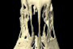
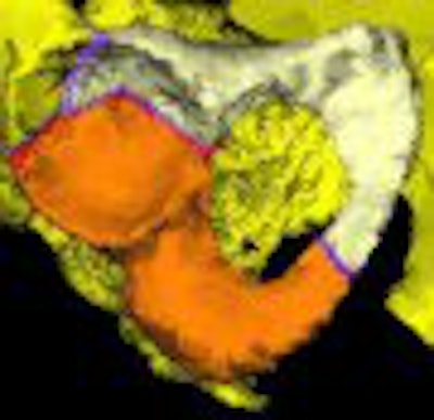
| Acetabular fracture fragments can be segmented and color-coded in both 3D (above) and 2D (below). Consequently, the observer can easily identify the control and result of the achieved reduction in 3D and 2D. |
 |
In an e-mail interview with AuntMinnie.com, co-author Dr. Daniel Kendoff said that at the time of the study the group was working on developing the module with a vendor, but that project is currently on hold. However, BrainLab of Feldkirchen, Germany, does market a similar 3D planning system, he said.
Five orthopedic trauma surgeons and two medical students performed planning and reduction trials on four plastic pelvis models. When the operator had determined that the reduction was satisfactory, an Iso-C3D scan was done to assess the final results. Segmentation of each fracture fragment was then mapped to a 3D coordinate system. Reduction accuracy was measured as an angular and linear displacement of each fragment relative to its anatomic position on the pelvic CT scan.
 |
| 3D imaging offers visual feedback of the operative result that can be corrected in the same intraoperative session. The overall judgment of the complex anatomy of the pelvis becomes more reliable for the surgeon, compared to any 2D visual feedback. Images courtesy of Dr. Daniel Kendoff and Dr. Musa Citak. |
According to the results, using the virtual planning module, the average translational malreduction of the fragments was significantly less (6.0 ± 4.7 mm) compared to conventional methods (9.2 ± 11.2 mm). The average reduction error following virtual planning was 11.6 ± 10.6° compared to 14.7 ± 10.7°.
The time for reduction was also significantly reduced. For T-type fractures, the average time with the virtual module was four minutes and 15 seconds, nearly three minutes less than with conventional methods. For both spinal column fractures, the time with the virtual simulator was three minutes versus six and a half minutes.
The authors noted that reductions were more accurate when done by the orthopedic trauma surgeons, but that a virtual simulator can be used as a training tool for young surgeons.
By Shalmali Pal
AuntMinnie.com staff writer
December 24, 2007
Related Reading
MRI, CT prove their metal for imaging orthopedic hardware, May 4, 2007
The ABCs of MRI for hip disorders, February 2, 2007
3D penetrates trauma imaging niche, August 4, 2006
Copyright © 2007 AuntMinnie.com