A new 3D intraoperative imaging technology could provide surgeons with submillimeter spatial resolution and soft-tissue visibility in near real-time, according to its creators at the University of Toronto (UT) in Ontario, Canada. Currently used in trials with human subjects, the conebeam CT on a mobile C-arm (CBCT C-arm) can acquire a full volume image in a single rotation.
Jeffrey Siewerdsen, Ph.D., from the University of Toronto, presented an overview of the CBCT C-arm at the 2007 American Association of Physicists in Medicine (AAPM) meeting in Minneapolis. Siewerdsen and colleagues, along with engineers at Siemens Medical Solutions of Malvern, PA, began collaborating in 2000 to find medical applications for Siemens' PowerMobil C-arm unit.
 |
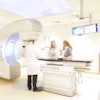
| Above, illustration of the mobile isocentric C-arm modified to include a flat-panel detector, motorized orbit, geometric calibration, and control system for CBCT acquisition. Below, intraoperative CBCT reconstructions provided by the host PC are displayed on an in-room display. |
 |
The CBCT C-arm is currently in trials with patients undergoing brachytherapy, as well as chest, breast, spine, and head and neck surgery at UT's University Health Network.
The CBCT C-arm combines three technologies: CBCT reconstruction, flat-panel detectors, and the isocentric (all x-ray beams have a central focus point) C-arm. The C-arm serves as the platform for image acquisition, and maintains a fixed field-of-view as the C-arm gantry moves around the patient. The mobility of the C-arm makes it applicable for emergency rooms and intensive care units, according to the group.
Siewerdsen said the CBCT C-arms scans at a lower dose than a standard diagnostic CT, making it safe to repeat imaging during a procedure. According to Siewerdsen, the typical doses for a head scan are 100 kVp (with added filtration to reduce beam-hardening effects) and 50 mAs resulting in about 3 mGy (0.1 mSv).
 |
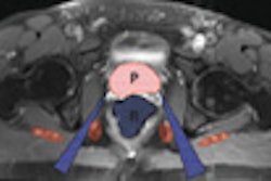
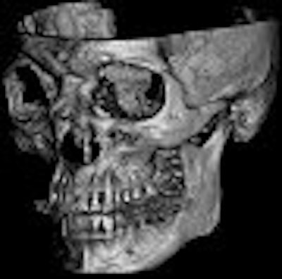
| The example image (skull) illustrates the 3D spatial resolution and FOV. Images courtesy of Jeffrey Siewerdsen, Ph.D., University of Toronto. |
While the researchers acknowledge that low resolution is a drawback of the CBCT C-arm prototype, the images should still be adequate enough to guide surgeons and radiologists to soft-tissue targets and critical structures. Imaging updates taken during the procedure could reveal any residual tumor remaining after excision, Siewerdsen said.
Previously assessed in phantom models and cadavers, CBCT C-arm images provided soft-tissue visibility (about 20 HU) and high spatial resolution (about 1 mm) at doses low enough (about 10 mGy) to allow repeat intraoperative imaging. Siewerdsen and colleagues also reported that the best images of soft tissue and bony detail were achieved with dynamic-gain readouts across all doses, whereas fixed gain was degraded at high doses because of pixel saturation, and image noise degraded the dual-gain readouts.
By Kathlyn Stone
AuntMinnie.com contributing writer
September 17, 2007
Related Reading
Chest CT a handy adjunct to breast MR, July 11, 2007
CARS news: Automated device tracking beats conventional CT/US guidance, June 28, 2007
Copyright © 2007 AuntMinnie.com