CHICAGO - A pilot study that combined transesophageal magnetic resonance imaging (TE MRI) with standard cardiac MRI for assessment of aortic plaque found the paired approach to be an excellent diagnostic option. Moreover, the technique is helpful for evaluating the efficacy of medical treatment, said lead investigator Dr. Henning Steen, a research fellow at Johns Hopkins Medical Institutions in Baltimore.
Steen said the dual technologies are so effective that "we are now using it to evaluate both aortic plaque and carotid plaque." He added that an ongoing study indicates that "by adding contrast we can also identify the composition of the plaque, not only calcification but the friability of the plaque. We can distinguish ‘hot’ plaque from ‘cold’ plaque." Steen presented his group’s results on Tuesday at the American College of Cardiology (ACC) meeting.
In an interview with AuntMinnie.com, Steen said the paired technology is now being used to "evaluate the effect of different statin therapies. We get baseline images and follow up at 6 and 12 months to see if there is regression or progression of plaque."
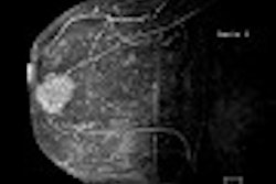
As with real estate, the benefit that TE MRI adds to aortic imaging boils down to location, location, location. With standard MRI, the coil gets no closer than 6 or 10 cm, which means that there is "so much noise that part of the signal is lost. With TE MRI we can place a coil much closer to the aorta, which cuts down on noise and improves the signal so the resolution is better," he said. The TE MRI receiver is threaded on a nasogastric route into the esophagus.
In the pilot study, 19 subjects were evaluated with 228 images of the thoracic aorta obtained with both surface and TE MRI coils using a 1.5-tesla magnet. The array surface coils were positioned anteriorly and posteriorly on the chest, coupled with a specially designed TE receiver coil positioned in the esophagus through a small-caliber nasogastric tube.
Fast spin-echo with inversion recovery and proton-density (PDW) and T2-weighted sequences were used for atherosclerotic plaque imaging. Signal-to-noise ratio (SNR) of atherosclerotic plaque in the descending thoracic aorta, ascending aorta, and aortic arch was calculated for the combined approach and for surface and TE MRI coils individually by two independent observers.
The SNR of the combined surface and TE MRI approach was superior to that of individual coils 93% of the time, Steen said.
In the descending thoracic aorta (180º arc) close to the TE coil, the TE coil contributed 68% of the combined SNR for T2-weighted images (95% confidence interval, 57%-78%). The surface coils accounted for 32% (22%-43%) of combined SNR.
In the descending thoracic aorta, remote from the TE coil, the TE coil contributed 37% (26$-48%) of combined SNR for T2-weighted images. The SNR contribution from the surface coils was 63% (52%-73%) in this region.
In the aortic arch, the TE coil SNR was 63% (53%-72%) of combined SNR for T2-weighted sequences. In the ascending aorta, the TE coil contributed <25% to the combined SNR for T2-weighed images. SNR values for PDW images were similar to T2-weighted images.
Steen said that Hopkins is currently planning "a much larger study so that we can demonstrate clinical utility."
By Peggy PeckAuntMinnie.com contributing writer
April 4, 2003
Related Reading
Calcified plaque increased in coronary arteries of diabetics, March 20, 2003
MRI links carotid artery plaque inflammation to unstable angina, March 10, 2003
Thin-collimation MDCT identifies more coronary calcium, December 18, 2002
Copyright © 2003 AuntMinnie.com