Women who carry the BRCA1 or BRCA2 genetic mutation are eight times likelier than the general population to develop breast cancer. However, a clear-cut explanation of why this increased risk exists is still under investigation. One theory is that these women exhibit a higher breast density. This increased density reflects a larger amount of tissue, and as most breast cancers develop from epithelial cells that line the breast ducts, a greater amount of tissue could mean a greater chance of developing cancer.
Researchers from multiple institutions tackled this issue by using computer image analysis of digitized mammograms from BRCA1 and BRCA2 mutation carriers. They looked at mammographic parenchymal patterns as well as density in these women, comparing them to images of women at low risk for breast cancer. Their results were published in Radiology (August 2002, Vol. 224:2 pp.560-568).
"To be eligible for the study, mutations carrier must have had mammograms obtained after 1990 and before cancer was diagnosed. A total of 30 BRCA1 and BRCA2 mutation carriers had mammograms that could be used for evaluation," wrote lead author Zhimin Huo, Ph.D., from the University of Chicago. Huo’s co-authors were based in Chicago, as well as at the University of Pennsylvania in Philadelphia.
The 142 low-risk patients could not have a family history of breast cancer. And based on Gail model estimates, their lifetime risk of developing breast cancer had to be less than 10% to participate in the study.
The mean age of the genetic mutation carriers was 42.7 years; the mean age of the low-risk women was 44.7 years. In order to rule out possible age-related bias, 60 low-risk individuals were matched at 5-year intervals with 30 mutation carriers.
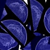
Mammograms were digitized with a laser scanner (LD 4500, Konica Medical Imaging, Wayne, NJ) at 0.1-mm pixel size and 10-bit gray level. Regions of interest (ROI) of 256 x 256 pixels were manually selected from the central breast region, immediately behind the nipple, because this area usually includes the densest parts of the breast.
Using linear discriminant analysis (LDA), skewness, balance, contrast, and coarseness were selected as the computer-extracted features used to evaluate the mammographic characteristics of an extended group. Receiver operating characteristic (ROC) analysis was used to determine the usefulness of individual computer-extracted features in differentiation between the two groups.
The usefulness of the individual features, as well as of LDA, was indicated in terms of Az values, or area under the ROC curve.
"A dense ROI should yield a negative value of skewness, whereas a fatty ROI should yield a positive value of skewness," the authors stated. "As characterized by the computer-extracted features, the mutation carriers tended to have more dense breast tissue than did the low-risk women…and their mammographic patterns tended to be coarser in texture and lower in contrast than those in the low-risk cases."
Based on LDA, the Az values were 0.91 and 0.92 in distinguishing between the BRCA1 and BRCA2 mutation carriers and low-risk women. The estimated standard of error for the two Az values was 0.03, the authors reported. The texture feature of coarseness yielded the highest Az value among all four features.
In addition, "the density rating distribution of the 30 BRCA1 and BRCA2 mutations carriers and the 60 age-matched low risk cases shows that the low-risk women tended to have fatty breasts when compared with the 30 mutations carriers," the authors wrote. "BRCA1 and BRCA2 mutation carriers tended to have more dense breast tissue, and their mammographic patterns tended to be coarser, with lower contrast than those of low-risk women."
The researchers concluded that analyzing digital mammograms with multiple computer-extracted features yielded better performance in differentiating between low-risk women and mutation carriers.
"Furthermore, our objective computerized method enables characterization of the mammographic patterns associated with breast cancer risk, and may aid estimation of an individual’s risk of developing breast cancer," they concluded.
By Shalmali PalAuntMinnie.com staff writer
October 25, 2002
Related Reading
Effect of family history on breast cancer survival varies, June 14, 2002
MRI successfully tracks women with hereditary breast cancer risk, April 8, 2002
MRI superior to mammography in screening high-risk women for breast cancer, July 27, 2001
Copyright © 2002 AuntMinnie.com