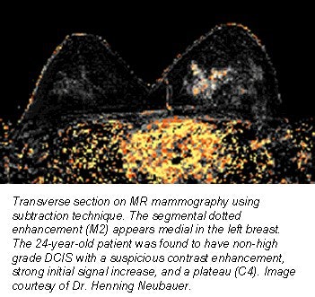
MR mammography turns in a solid performance when it comes to high-grade ductal carcinoma in situ (DCIS). However, it may be less effective for smaller, non-high-grade DCIS lesions, according to German investigators. In a presentation at the 2001 RSNA conference in Chicago, Dr. Henning Neubauer from Friedrich Schiller University in Jena discussed his group’s work on MRI's sensitivity and morphological enhancement patterns for DCIS.
"In our study, we wanted to answer the following questions: Is there any possible increase in signal and time intensity curves on dynamic MRI for DCIS? Is there a characteristic pattern of enhancement that we can identify?" said Neubauer.
For the study, 39 consecutive patients with pure DCIS underwent a routine MR examination on a 1.5-tesla Gyroscan S15 ACS II (Philips Medical Systems, Bothell, WA). Contrast-enhanced, T1-weighted, 2-D FFE images were obtained and then retrospectively evaluated.
"In order to classify our findings, we defined categories," Neubauer said. "One set of categories for signal increase, another set of categories for signal enhancement."
Signal increases were divided up as follows:
- C1= non-significant
- C2 = slow-continuous
- C3 = strong initial and slow further increase
- C4 = strong initial increase followed by a plateau phenomenon
- C5 = same as C4 with the addition of a washout phenomenon
Morphological patterns were assessed as:
- M0 = no pattern observed
- M1 = linear or linear-branched
- M2 = segmental dotted or granular
- M3 = segmental homogenous
- M4 = focal spot
A strong initial increase of at least 90% in the first two minutes of the exam (C4 and C5) was considered suspicious for malignancy, Neubauer said. The strong initial increase with additional slow enhancement (C3) also was found for benign tumors. All cases were correlated with histology.
 |
"The hallmark for DCIS as found in our study was segmental unilateral enhancement (82%) with a 'malignant' time-intensity curve in two-thirds (of the cases), and an intermediate signal increase in one-third of the cases," Neubauer wrote in an e-mail to AuntMinnie.com. "The assessment of the morphology revealed a dotted, granular pattern to be the predominant pattern of enhancement (73%)."
In the case of low-grade tumors, only 15% showed a wash in pattern, but no washout, while 8% showed slow enhancement. However, on the majority of MR exams, there was "no enhancement. We just didn’t see them," Neubauer said during his presentation.
This led the group to conclude that MR mammography is sensitive enough for high-grade DCIS, yet of little value for smaller tumors.
"We think it absolutely necessary to perform the routine MR mammography with bilateral scans (unlike the unilateral scans commonly done in the U.S.). Only the evaluation of both breasts in the same scan makes it possible to exclude the most important differential diagnosis -- that is enhancement due to hormone effects with a typical bilateral enhancement (while DCIS is unilateral, as a rule)," Neubauer wrote.
According to another RSNA presentation, hormone replacement therapy (HRT) can influence the signal intensity in dynamic MR mammograms. Dr. Steffen Sachse and co-authors, also from Friedrich Schiller University, studied the MR mammography results of 201 patients who were on HRT. The majority of patients, 85%, showed "bilateral, symmetrical, synchronous, progressive enhancement, which were partially confluent on late scans." However, the group did not encounter the plateau phenomenon or a washout effect.
By Shalmali PalAuntMinnie.com staff writer
January 2, 2002
Related Reading
Patients with known breast malignancy benefit from MRI, November 25, 2001
MRI superior to mammography in screening high-risk women for breast cancer, July 27, 2001
Hormone replacement therapy complicates breast imaging, June 11, 2001
Copyright © 2002 AuntMinnie.com


