The most common reason for ultrasound of the thigh at our institute, the Studio Ecograficao Dott. Stefano Ciatti in Prato, Italy, is related to muscle injury, usually resulting from sporting or recreational activities that require sprinting or jumping.
The rectus femoris muscle is composed of fibers appropriate for rapid, forceful activity and crosses two joints (the hip and the knee). These characteristics may explain why this muscle is particularly vulnerable to eccentric stress forces, such as those resulting from the powerful contractions associated with sprinting and jumping. Soccer and rugby are the sports most commonly associated with injury to this muscle in the patients who visit our institute.
Clinical history
The patient is a 17-year-old soccer player who injured himself while slowing to kick the ball during a competitive game. He slipped while performing this movement and experienced immediate pain in his right thigh and hip.
Physical examination
The patient presented with a painful and tender lump located at the middle third of the anterior thigh the day after his injury. No plain radiographs had been performed.
Discussion
The rectus femoris muscle tends to tear at the level of its proximal or distal myotendinous junction rather than within the muscle belly. Tears affecting the distal insertion of the muscle fibers into the deep distal aponeurosis seem more common. However, proximal tears are increasingly detected, as demonstrated by recent articles reporting on their MR and ultrasound imaging appearances.
Although MR imaging has been described for evaluation of rectus femoris tears, ultrasound is valuable to appreciate the size of the rupture, rule out other conditions, allow longitudinal assessment, and eventually guide the evacuation of the hematoma. Rectus femoris tears affecting the deep distal aponeurosis are frequently detected based on clinical findings.
Athletes with acute tear of the deep distal aponeurosis present with a painful lump located at the level of the middle third of the anterior thigh corresponding to the retracted rectus femoris muscle. Active flexion of the thigh associated with extension of the leg causes the anterior mass to become more evident due to muscle contraction.
Partial avulsions are recognized as focal fusiform hypoechoic areas within the muscle. Longitudinal ultrasound images are well suited for evaluating deep distal aponeurosis tears and clearly demonstrate the retracted fibroadipose septa lying at a variable distance from their normal anatomical site of attachment into the aponeurosis.
The gap caused by the tear may be filled with fluid or hypoechoic tissue related to hemorrhagic infiltration. Complete tears of the rectus femoris with detachment of the muscle from the quadriceps tendon are more frequently detected with ultrasound. These appear as a complete separation of the muscle belly, which assumes a rounded and irregular distal shape instead of its classical pointed end. Acute cases demonstrate a hypoechoic blood collection distal to the torn muscle and superficial to the thickened and irregular deep distal aponeurosis.
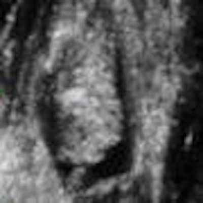
Initially, the examination of this patient was performed using traditional 2D ultrasound and subsequently with 3D ultrasound. Longitudinal scanning demonstrated the most distal portion of the rectus femoris muscle retracted with a swollen blunt end as a result of avulsion (Fig. 1). Active flexion of the thigh associated with extension of the leg causes the anterior mass to become more evident due to muscle contraction; this was observed in real-time ultrasound scanning. However, in order to permit patient comfort, 3D volume acquisition was performed with the patient in a relaxed position.
 Fig. 1: Longitudinal plane demonstrating a distal aponeurosis tear. Note the retracted rounded end of the muscle and the presence of a distal hematoma (black area). All images courtesy of Dearbhla O'Dwyer and Dr. Stefano Ciatti.
Once the location of the tear was determined, we decided to perform a 3D volume acquisition to better study the condition of the muscle and the full extent of the tear. Three-dimensional volume acquisition permits one to scroll backward and forward and up and down through the acquired volume.
The coronal plane permits a frontal view traditionally unavailable in ultrasound scanning that frequently allows a better appreciation of the extent of a tear (Fig. 2). As with many modern imaging modalities, 3D reconstruction is used to demonstrate both normal and abnormal anatomy. In this particular case, the complete separation of the muscle fibers from the point of insertion is well demonstrated along with a large hypoechoie fluid collection and coagulation material with the use of the coronal plane and 3D reconstruction (Fig. 3 and 4).
 Fig. 2: Coronal plane clearly demonstrating the retracted muscle fibers with a swollen blunt end as a result of avulsion and surrounding hematoma. We noted a small quantity of coagulation material distally and deep to the tear.
 Fig. 3: Using multiplanar imaging and 3D reconstruction, it is possible to confirm the presence of the muscle tear at the level of the distal aponeurosis surrounded by hematoma or hypoechoic tissue related to hemorrhagic infiltration and coagulated material.
 Fig. 4: 3D reconstruction (enlarged image) demonstrating the intact vastus intermedius and vastus lateralis with the complete rupture of the central rectus femoris muscle. Note the black fluid collection and hematoma distally with coagulated material beneath the retracted swollen muscle.
Twenty days after this initial ultrasound examination, the patient presented for a checkup scan. He was feeling much better and no longer suffered from pain. His trainers were keen to get him back to training. Once again, the muscle was examined with both 2D and 3D ultrasound.
A new volume acquisition was obtained and studied to perform a detailed examination of the condition of the muscle and assist in a complete assessment of the patient's condition before putting the muscle under new strain. The volume acquisition obtained demonstrating the condition of the muscle is shown below (Fig. 5 and 6).
 Fig. 5: Multiplanar imaging shows absorption of some of the hematoma but demonstrates the presence of the muscle tear.
 Fig. 6: 3D reconstruction (enlarged image) confirming the continued presence of the muscle tear and hematoma.
Healing of the tear appears as irregular hyperechoic tissue located distal to the severed muscle due to local scarring tissue. Volume loss and fatty infiltration of the injured rectus femoris reflecting post-traumatic atrophy may become evident. It is evident in this case that the muscle requires further time to heal, and the patient was advised to continue resting.
There is evidence of reabsorption of the hematoma and coagulated material that were present on the original volume acquisition. There was less fluid surrounding the torn muscle on the follow-up scan compared with the original scan, and it is clearly evident that the healing process requires more time. However, classical signs of irregular hyperechoic muscle tissue that is present with the healing process are absent. This patient did not visit our institute for any additional follow-up scans.
Conclusion
Ultrasound is a relatively low-cost imaging modality that readily permits examination of soft tissues, including muscle fibers. It allows an accurate assessment of the rectus femoris muscle, including its three aponeuroses. The superficial proximal and the deep distal aponeuroses appear as thickening of the anterior and posterior muscle fascia, respectively. On transverse ultrasound images, the central aponeurosis appears as a comma-shaped, sharply delineated hyperechoic band.
Ultrasound, including the use of multiplanar imaging and 3D reconstruction, gives valuable information as to the condition of muscle ruptures. Simultaneous representation of muscle tears under observation in three orthogonal planes allows better documentation and evaluation of their extent.
The coronal plane, unavailable in traditional ultrasound scanning, provides a plane from which one can better appreciate the extent of these tears. The use of automatic volume acquisition allows calculation in three orthogonal planes, and therefore permits accurate evaluation of the size of a tear.
Ultrasound can exclude other conditions, may be useful in aspiration of a hematoma, and can be used, as in this case, to follow up the post-traumatic reabsorption of a hematoma and reorganization of the muscle fibers. The assessment of the condition of the muscle fibers throughout the healing process can be controlled as the hematoma is replaced with irregular hyperechoic tissue located distal to the severed muscle caused by scar tissue. Volume loss and fatty infiltration of the injured rectus femoris reflecting post-traumatic atrophy are readily assessed using multiplanar ultrasound.
By Dearbhla O'Dwyer and Dr. Stefano Ciatti
AuntMinnie.com contributing writers
June 16, 2008
Sonographer Dearbhla O'Dwyer, B.Rad, H.Dip D.I. (U/S), and Dr. Stefano Ciatti are in private practice at the Studio Ecograficao Dott. Stefano Ciatti in Prato, Italy.
Related Reading
Pictorial essay: 3D ultrasound reveals shoulder pathology, March 21, 2008
US guidance needles away the pain of rotator cuff calcific tendinitis, November 29, 2007
Making the most of MRI to assess the rotator cuff pre- and postinjury, November 9, 2007
US shows full spectrum of shoulder injuries, September 26, 2006
References
Bianchi S, Martinoli C. Ultrasound of the Musculoskeletal System. Springer-Verlag, Berlin, 2007.
Bianchi S, Martinoli C, Waser N, et al. Rectus femoris central tear. Skeletal Radiology, October 2002, Vol. 31:10, pp. 581-586.
Garrett WE Jr. Muscle strain injuries: Clinical and basic aspects. Medicine & Science in Sports & Exercise, August 1990, Vol. 22:4, pp. 436-443.
Lee JC, Healy J. Sonography of lower limb muscle injury. American Journal of Roentgenology, February 2004, Vol. 182:2, pp. 341-356.
Copyright © 2008 Dearbhla O'Dwyer and Dr. Stefano Ciatti