Differentiating benign from malignant lesions with contrast-enhanced ultrasound (CEUS) is best done at late phase, and a quantitative analysis of ultrasound video intensity can be used to calculate the conspicuity index for effective and objective differentiation of the liver tumors, according to Italian researchers.
Specifically, the researchers examined contrast enhancement and conspicuity after the injection of a sulfur hexafluoride-filled microbubble agent using two methods: visual analysis by three observers and quantitative analysis of the video intensity by digital means.
The retrospective study was conducted by Dr. Emilio Quaia and colleagues from Cattinara Hospital, University of Trieste in Trieste, Italy. Quaia presented the results at the 2004 RSNA meeting in Chicago.
The purpose was to assess if ultrasound video-intensity analysis after injecting the microbubble agent improved diagnostic performance in characterizing focal liver lesions. Quaia also referred to earlier studies that showed use of sulfur hexafluoride contrast led to improved characterization of solid focal liver lesions (European Radiology, June 2004, Vol. 14:6, pp. 1092-1099; Radiology, August 2004, Vol. 232:2, pp. 420-430).
Nonlinear low-acoustic imaging in pulse-inversion mode or contrast-tuned mode was performed on 179 patients after a 2.4-4.8 mL bolus injection of SonoVue (Bracco Diagnostics, Princeton, NJ). The lesions selected for the study were indeterminate at baseline ultrasound and color Doppler ultrasound, and ranged in size from 1-8 cm.
There were 101 malignant lesions, including 52 hepatocellular carcinomas and 78 benign lesions, in the group that were confirmed on histology, CT, or MR. Three onsite sonologists recorded their observations separately after visual analysis.
Depending on the conspicuity, different grades were assigned to the lesions. Hypovascularity was graded from -2 up to 0, with 0 indicating an isovascular lesion. Hypervascularity was graded from 0 up to +2.
The findings of the visual analysis showed there was an overlap between the benign and malignant lesions in the arterial phase. However, in the late phase, benign lesions usually presented as isovascular compared to malignant lesions, except in some instances in which malignant lesions also presented as benign with an isovascular appearance, Quaia said.
Quantitative video-intensity analysis was used to remove subjectivity and correct the mismatch between observer readings. "One observer performed objective assessment on four to eight digital 8-bit images in tagged-image format (TIFF) selected from arterial (10-35 secs) and late (95-180 secs from microbubble injection) phase," the group explained in their RSNA abstract.
Three TIFF frames each at arterial phase and late phase were used for the quantitative analysis. The grayscale intensity within the lesion was computed and compared with the intensity in the adjacent liver parenchyma.
The intensity outside the liver was kept constant in each case at 35-40 grayscale level. "In each image two equal (1500-4187 pixels, mean 1965 pixels) rectangular regions of interests were drawn, the first to encompass the lesion and the second the adjacent liver parenchyma," they wrote. The grayscale intensity was measured on a scale from 0 (black pixels) to 255 (white pixels).
The objective conspicuity index (OCI) was calculated by comparing the intensity within the lesion to the intensity in the adjacent liver parenchyma and localizing it with the liver intensity.
 |
 |
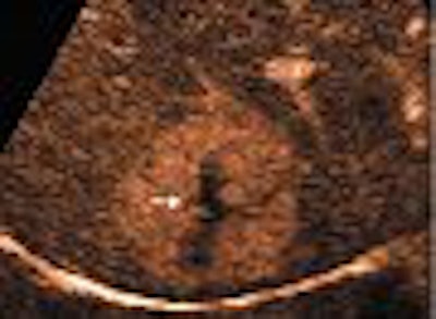
| Transverse contrast-enhanced US scans obtained in contrast-specific mode with pure harmonic detection in 45-year-old woman show typical spoke wheel-shaped contrast-enhancement pattern of focal nodular hyperplasia after microbubble contrast agent injection. Above, central spoke wheel-shaped contrast enhancement (arrow) is evident 15 seconds after the injection (arterial phase). Middle, contrast enhancement (arrow) is diffuse and homogeneous 25 seconds after the injection (arterial phase). Below, contrast enhancement is hyperechoic 105 seconds after the injection, with a central hypoechoic region that corresponds to the central scar (arrow) that is evident during the late phase. |
 |
| Quaia E, Calliada F, Bertolotto M, et al, "Characterization of Focal Liver Lesions with Contrast-specific US Modes and a Sulfur Hexafluoride-filled Microbubble Contrast Agent: Diagnostic Performance and Confidence," Radiology 2004;232:420-430, figure 8a-c. |
The OCI was equal to 0 for isovascular lesions, less than 0 for hypovascular lesions, and greater than zero for hypervascular lesions. The difference between benign and malignant lesions was more pronounced in the late phase. Malignant lesions presented a predominantly hypovascular appearance compared to benign lesions, which presented hypervascular appearance.
The researchers observed that hepatocellular carcinoma was hypovascular at late phase but could, however, also present as hypervascular or isovascular, at times. "Other malignant lesions are almost completely isovascular," Quaia said.
An objective contrast enhancement score (OCES) was also calculated with the difference in lesion intensity before and after contrast injection and the difference in liver intensity before and after contrast injection, as parameters.
The difference between the median OCES and OCI values was assessed for malignant and benign lesions. The values were not significant at the arterial phase (p > 0.05), but were significantly different at the late phase (p < 0.05).
"In particular, OCI revealed a value less than 0 (median -0.4) in 73/95 (76%) malignant lesions and a value greater than 0 (median +0.65) in 48/52 (92%) benign lesions. Considering OCI value at late phase to differentiate benign from malignant lesions (less than 0 = malignant; greater than 0 = benign), overall accuracy (± confidence interval) corresponded to 82 ± 0.06%," the authors wrote.
By N. Shivapriya
AuntMinnie.com contributing writer
March 29, 2005
Related Reading
Contrast US pinpoints focal liver lesions, results in cost-savings, March 25, 2005
Contrast US tops conventional US for blunt liver trauma, March 21, 2005
Proton magnetic resonance measures liver iron levels noninvasively, February 3, 2005
Early arterial phase best for HCC detection, December 28, 2004
Copyright © 2005 AuntMinnie.com


