(Ultrasound Review) 3-D ultrasound measurements of the common bile duct (CBD) correlated well with 2-D measurements in a study by Dr Ashutosh Rao and colleagues, published in Journal of Ultrasound in Medicine. They established that 3-D ultrasound was an accurate and reliable method for evaluating the CBD size.
"A prospective blinded study was initiated to compare measurements obtained from traditional 2-D examinations versus 3-D sonography," they wrote.
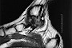
The group from the University of Texas Southwestern in Dallas examined 55 consecutive patients who presented for abdominal ultrasound for a variety of reasons. Two patients had previous cholecystectomy. Both 2-D and 3-D imaging was performed using a 3.0-5.0 transducer.
Initially, 2-D anterioposterior (AP) measurements were made in the long axis and then a 3-D acquisition was performed of the right upper quadrant with the breath held for five seconds. Three-dimensional data sets were electronically stored for subsequent evaluation.
According to the authors, "Our study shows no significant differences in AP diameters measured in 3-D long axis versus 3-D short axis views, and these measurements in fact had a high degree of correlation."
They added, "This finding would be expected, given that the same structures are being measured in the same place (i.e., the anterior and posterior common duct walls), and is a validation of the accuracy of measurements between projection planes in 3-D sonography."
Three-dimensional ultrasound only added one minute to the overall scanning time, but enabled better visualization of the CBD in uncooperative patients and patients with complicated anatomy. "Advantages of 3-D sonography of the common duct include rapid acquisition of data that can be processed and viewed at a later time and increased confidence in the common duct diameter measured," they wrote.
The 2-D and 3-D measurements were not exactly the same but showed a correlation. The mean 2D AP long axis diameter was 3.6 mm, and the mean 3D AP long axis and short axis diameters were 4.1 mm.
"The two-dimensional common duct measurement correlated with the long axis anteroposterior three-dimensional measurement (P < .001), the short axis anteroposterior three-dimensional, and the short axis transverse three-dimensional measurement," they concluded.
Three-dimensional sonographic evaluation of the common bile ductRao, A. V., et al
Department of radiology, University of Texas Southwestern, Dallas.
J Ultrasound Med 2003 September; 22:939-944
By Ultrasound Review
October 29, 2003
Copyright © 2003 AuntMinnie.com