Fetal cardiac screening, done between 13-14 weeks’ gestation and again at 22 weeks’ gestation, should be performed as a matter of course in order to catch cardiac anomalies in time, according to Dr. Reuben Achiron from Tel Aviv University School of Medicine.
"Give me one good reason why everyone is not doing this," said Achiron, an obstetrician who specializes in obstetrical ultrasound. "Fetal cardiac scans are often only done if there is a specific indication, but we believe that the heart is part of the fetal body and should be part of the regular scan."
Achiron, who also is chairman of obstetrics and gynecology at Sheba Medical Center in Ramat Gan, Israel, said that in urban areas of his country, about 80% of women have early fetal cardiac screening done as part of their routine care.
"There is a big demand for this, patients are asking for it -- they know that it is possible and they want it done," he said.
Speaking at the 2003 congress of the World Federation of Ultrasound in Medicine and Biology (WFUMB) and the American Institute of Ultrasound in Medicine (AIUM), Achiron explained that 64% of fetal cardiac anomalies can be spotted in an early scan, using high-resolution transvaginal ultrasonography.
"Many of the cardiac malformations, about 50%, are associated with other chromosomal anomalies," he said. "For example, 50% of Down’s syndrome patients have a cardiac defect. So you have a chance of detecting half of Down’s syndrome babies just by looking at the heart. If a woman is young and she is not planning to have an amniocentesis, the early fetal cardiac screen is very valuable."
Achiron said the early transvaginal fetal cardiac exam is based on five echographic planes:
- The first and most caudal plane is a transverse view of the upper abdomen.
- Moving cephalid, there should be a traditional four-chamber view, which remains key to the fetal echocardiographic examination.
- There should be a plane introduced to visualize the outflow tracts of both ventricles.
- A plane to view the arches of the ductus and the aorta.
- There should be a transverse mediastinal view including the three vessels and the trachea.
Another advantage of the early scan is to identify malformations that can be treated in-utero. And even in the case of malformations that can only be treated postnatally, early diagnosis remains very important.
 |
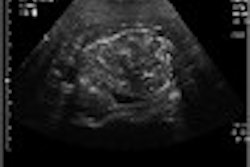
| The fetal cardiac scan depicts the heart at 14 weeks’ gestation (8 mm). More than half of fetal cardiac anomalies can be spotted in an early scan, using high-resolution transvaginal ultrasonography. Image courtesy of Dr. Reuben Achiron. |
"If you detect problems in utero, the prognosis is much better in terms of morbidity and mortality. Patients know that they should deliver in specialized centers, and the system is prepared before the delivery," Achiron explained.
Achiron said sonographers, or other physicians, who are accustomed to performing fetal echocardiograms later in gestation should not find it difficult to orient themselves in the smaller fetal heart. However, his personal preference is that obstetricians perform the scans.
"This is a new field, fetal medicine, but I think the obstetrician is the care provider for the fetus and this is part of the care they provide. They will have to learn the technique, but it is not that different from what they already know," he said.
The second fetal cardiac scan, at 22 weeks’ gestation, is important for catching such anomalies as aortic and pulmonic stenosis, cardiac rhabdomyoma, tetralogy of Fallot, aortic coarctation, sealed foramen ovale, ventricular septal defects, and hypertrophic cardiomyopathy, which can only be identified later in pregnancy.
In a related presentation, Dr. Ilan Timor-Tritsch, professor of obstetrics and gynecology at New York University School of Medicine, supported the second scan. Based on his research, early fetal cardiac imaging remains a particular challenge for sonographers.
He presented preliminary data on seven sonographers at his institution who performed transvaginal ultrasonography on 223 women between 11 and 14 weeks’ gestation. Adequate visualization of cardiac structures was less than 40% for all views of the heart except the axis.
"A second transabdominal ultrasound is definitely necessary to complement the early scan," Timor-Tritsch said.
In the U.S., cardiac ultrasound is considered part of basic fetal screening as defined by the American College of Radiology (ACR), the American College of Obstetricians and Gynecologists (ACOG), and the AIUM. However, it is usually only done in the middle trimester, and only if there is an indication, according to Dr. Joshua Copel, professor of obstetrics/gynecology and pediatrics at Yale University School of Medicine in New Haven, CT.
In his commentary on the two studies, Copel suggested that nuchal translucency (NT) screening, which detects 60% of cardiac anomalies (independent of those associated with chromosome abnormalities), would be a more cost-effective method of fetal cardiac screening, instead of the standard four-chamber heart screen.
"The problem is, relatively few people in the U.S. are trained to do the NT screen in the proper way. But if they were trained, it would be less expensive for each diagnosis that is made," he said.
By Kate JohnsonAuntMinnie.com contributing writer
July 1, 2003
Related Reading
Umbilical artery Doppler has edge in fetal distress screening, June 19, 2003
Sonography reveals predictors of lethal skeletal dysplasias, May 31, 2003
Pediatric CT won't stop growing, but dose can be minimized, May 8, 2003
Tissue velocity imaging shows promise in detecting fetal arrhythmias, September 10, 2002
Copyright © 2003 AuntMinnie.com