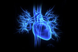
Combining myocardial scar and coronary images acquired with 3-tesla MRI into fused volumetric images can aid preprocedural planning for cardiac resynchronization therapy or coronary artery revascularization, according to a study in the September issue of the Journal of the American College of Cardiology: Cardiovascular Imaging.
A study team from the University of Western Ontario's Robarts Research Institute in London, Ontario, reported that its fusion technique was feasible in a study of 55 patients preparing to receive these vascular-based interventions. The group also found that referring physicians deemed fused imaging to be clinically valuable in approximately two-thirds of the cases (JACC Cardiovasc Imaging, September 2010, Vol. 3:9, pp. 921-930).
"[Three-tesla] MRI allows for high-resolution 3D scar imaging to be performed in a relevant patient population, and this, in turn, can be fused to a matched 3D image of the coronary vasculature," lead author James White, MD, told AuntMinnie.com. "The inherent spatial registration of these images affords very rapid fusion into a single volumetric dataset."
White said the researchers pursued this approach to provide a more sophisticated and intuitive method for selecting optimal vascular targets for specific cardiovascular procedures.
The use of 3D CT vascular angiography can identify these targets, but it does not account for myocardial scar tissue, which has been shown to hinder patient response to some heart failure therapies. For example, investigators have found that scar in the region of where pacemaker leads are typically delivered during cardiac resynchronization therapy prevents clinical response, White said.
Simultaneous visualization of veins and scar as these leads are placed through the coronary veins would allow for selection of the most appropriate vein that is not associated with scarred muscle, he said. This ability would also be appropriate for selecting coronary arteries for bypass or angioplasty, as opening blood vessels to scarred muscle does not provide improvements in heart function, White said.
With this goal in mind, the researchers sought to develop a combined 3D imaging technique that spatially registers both myocardial scar and vascular anatomy. First, a 3D coronary MR image is obtained using continuous infusion of gadolinium contrast.
Because scar tissue retains the contrast agent after it has been washed out of the blood stream and normal tissue, a repeat 3D study using the same pulse sequences is then performed 20 minutes later to highlight the scar. The images are then fused together in a 3D model that shows both vessels and scar tissue, according to the researchers.
"Using the described technique, we are able to provide not only an attractive image, but a visually intuitive 3D model of the heart that can immediately provide this relevant information to the clinician," White said.
|
Having trouble viewing this clip? Click here to view full-size clip or to change format. |
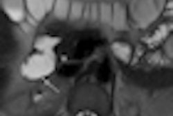
| 3D image of the heart. Video courtesy of Paul White, MD. |
|
Having trouble viewing this clip? Click here to view full-size clip or to change format. |
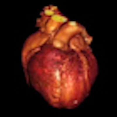
| 3D fusion images of coronary vasculature and myocardial scar tissue. Video courtesy of Paul White, MD. |
The researchers evaluated their technique in 55 studies in patients referred for either cardiac resynchronization therapy (42 patients) or coronary artery vascularization (13 patients). The mean age was 57 ± 14 years.
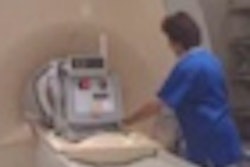
Both coronary-enhanced and scar-enhanced imaging were performed on a 3-tesla cardiac MRI scanner (Magnetom Trio, Siemens Healthcare, Malvern, PA). The same cardiac-gated, 3D, free-breathing, cardiac MRI technique was used during slow gadolinium infusion, as well as during the second scar-enhanced procedure 20 minutes afterward.
The matched datasets were then fused and volume-rendered to simultaneously display both coronary anatomy and myocardial scar. Using the open-source OsiriX software, an experienced, blinded observer performed visual scoring on a scale of 0 (noninterpretable) to 4 (excellent quality) to assess the image quality of the coronary artery, coronary vein, and myocardial scar.
The combined 3D coronary and scar imaging protocol was successful in 49 studies (89%). The presence of myocardial scar was found in 44 studies (80%) from 42 patients.
The researchers noted a quality score of 2 or greater in 97% of proximal- and midcoronary artery and vein segments. The 3D scar imaging technique produced a mean quality score of 2.8 ± 1.0 and was scored as 2 or greater in 86% of patients with myocardial scar.
Successful image fusion was achieved in all 36 patients with a scar imaging quality score of 2 or greater, according to the researchers. The researchers found underlying transmural scar below one or more planned vascular targets in nine patients (39%) set to receive cardiac resynchronization therapy and in eight patients (62%) targeted for coronary artery revascularization.
Referring physicians were provided with reports that identified which target vessel or vessels had underlying scar, as well as its transmural extent, according to the researchers. Representative 3D fused images were also included.
These referring physicians were then asked to score the clinical impact of the reports on their planned procedures on a scale of 0 (negative impact; led to confusion) to 4 (major impact; procedure cancellation). For the purposes of the study, a score of 2 or greater was considered to be a clinically significant impact.
The surveys showed an incremental clinical impact in 24 (67%) of the patients. In six patients, procedures were canceled because of the imaging results.
The clinical implications of the research remain to be explored; the next phase will examine how providing these images for clinicians during specific therapies changes their clinical success, White said.
"Should this prove to be a beneficial effect, this type of imaging may be useful in guiding the optimal delivery of vascular-based interventions where myocardial scar is recognized to be a significant obstacle to clinical response," White said.
By Erik L. Ridley
AuntMinnie.com staff writer
September 23, 2010
Related Reading
CT, MRI look at different etiologies for myocardial fat, September 25, 2009
New 3D cardiac MRI tool helps preplan pediatric surgery, August 12, 2009
Dobutamine CMR gauges cardiomyopathy treatment response, May 2, 2007
Copyright © 2010 AuntMinnie.com