Contrast-enhanced MRI dynamic evaluation of the bowel wall can effectively visualize inflammation caused by celiac disease, aiding the assessment of patients with this condition, according to a study in the September issue of Radiology.
Researchers from the University of Rome "La Sapienza" also concluded that an MRI exam could be useful in monitoring diet therapy and testing the use of anti-inflammatory drugs in patients with celiac disease. The lead author of the study was Gabriele Masselli, MD (Radiology, 2010, Vol. 256:3, pp. 783-791).
Celiac disease is one of the most common gastrointestinal diseases. The condition damages the small intestine and interferes with the absorption of food nutrients. People with celiac disease cannot digest gluten, which is a protein found in wheat, rye, and barley.
As the researchers noted in the study, MRI is becoming increasingly prevalent in the evaluation of patients with small-bowel diseases. The modality's ability to image soft tissue without ionizing radiation allows for repeated data acquisition over time without the patient being exposed to repeated doses of radiation in follow-up studies.
Patient enrollment
Researchers enrolled a total of 60 consecutive outpatients (21 men and 39 women) with a mean age of 27 years (range, 17-52 years), who were referred to the facility's gastrointestinal unit from February 2007 to July 2009 with symptoms of celiac disease.
During the two-year period, 45 patients (19 men and 26 women) already were diagnosed with celiac disease and were on a gluten-free diet for at least one year. To assess the reproducibility of the MRI measurements, 10 (22%) of the 45 patients who were treated for celiac disease underwent repeat MR imaging six to eight months later.
In addition, 30 patients (13 men and 17 women; mean age, 31 years) with familial polyposis syndrome and no history of celiac disease or other inflammatory small bowel diseases who also underwent MRI screening were included in a group of healthy control patients. All patients with untreated celiac disease underwent MR enterography within two weeks of gastroduodenal endoscopy.
For the MR enterography exam, patients fasted for 12 hours before undergoing MRI. Images were acquired with a 1.5-tesla MRI system (Magnetom Avanto, Siemens Healthcare, Erlangen, Germany) with an eight-channel abdominal phased-array coil. A polyethylene glycol (PEG)-water solution was used as an intraluminal contrast agent for small-bowel MR imaging.
Radiologist review
 |
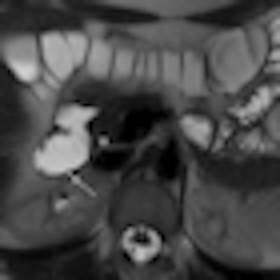
| 1.5-tesla MR images show a portion of the duodenum wall in a patient with untreated celiac disease (above), a patient with treated celiac disease (below), and a healthy patient (bottom). The images show no significant differences in duodenal wall thickness (arrow). |
 |
 |
The researchers calculated the mean maximum enhancement for each region of interest using a formula developed by Lavini and colleagues that takes into account maximum signal intensity and baseline signal intensity, and which assigned numerical values based on maximum enhancement.
Their analysis found that a mean maximum enhancement of the duodenal wall was 229.1 ± 46.4 in untreated patients with celiac disease, 109.8 ± 27.8 in treated patients with celiac disease, and 94.7 ± 17.9 in control subjects.
"This MR parameter was significantly higher in patients with untreated celiac disease than in patients with treated celiac disease and control subjects," the authors wrote, while maximum enhancement did not significantly differ between the patients with treated celiac disease and controls.
The researchers also found that duodenal wall thickness did not significantly differ between patients with celiac disease who were not treated for the condition, patients with celiac disease who were treated, and healthy control subjects.
There also was no significant difference between the maximum enhancement values determined at baseline and those determined six to eight months later in the 10 patients treated for celiac disease who underwent two MR examinations.
In conclusion, the results "suggest that dynamic evaluation of the bowel wall by using contrast-enhanced MR imaging can be an effective and reproducible way to show the inflammation state in celiac disease and useful in evaluating patients with this disease," according to Masselli and colleagues.
Given that their research was a single-institution study, the authors recommended that additional prospective multicenter trials be conducted "to clarify the clinical usefulness of dynamic contrast-enhanced MR imaging as a marker of therapeutic response."
By Wayne Forrest
AuntMinnie.com staff writer
September 1, 2010
Related Reading
Link seen between cognitive impairment and celiac disease, October 18, 2006
High resolution, fast acquisition give 3D MRA edge in abdomen, January 20, 2006
BMD measurement not required for most with celiac disease, November 17, 2005
Mitral valve prolapse tied to celiac artery stenosis, December 21, 2004
Broad range of neurologic disorders found in patients with celiac disease, June 22, 2004
Copyright © 2010 AuntMinnie.com