Canadian researchers have found that 1-tesla extremity MRI performs just as well as conventional 1.5-tesla MRI in evaluating hands and wrists of patients with rheumatoid arthritis, according to a study published in the June issue of Radiology.
A study team led by Dr. Ali Naraghi from the department of medical imaging at Toronto Western Hospital prospectively recruited 32 patients from a rheumatology outpatient clinic between December 2006 and November 2007 (Radiology, June 2009, Vol. 251:3, pp. 829-837). The study population included 30 women and two men, with a mean age of 52 years, who were diagnosed with rheumatoid arthritis.
The researchers included patients of any disease severity, activity level, and duration of symptoms, while patients with prior surgery to the affected extremity and those who chose not to undergo contrast-enhanced imaging were excluded from the study. Three patients who experienced claustrophobia and discomfort underwent limited examinations with the conventional 1.5-tesla scanner, but subsequently were excluded from the study.
Patient sample
The remaining 29 patients underwent complete MRI studies by both the 1-tesla extremity MRI system (MSK Extreme, ONI Medical Systems, Wilmington, MA) with a 100-mm v-SPEC coil (ONI) and a conventional 1.5-tesla system (Signa Excite II, GE Healthcare, Chalfont St. Giles, U.K.) with a dedicated eight-channel wrist coil (Precision Eight, Invivo, Orlando, FL).
Twenty patients received hand scans for their metacarpophalangeal joints, and nine patients had their wrists examined. Imaging was performed before and after intravenous injection of 0.1 mmol/kg of gadodiamide (Omniscan, GE Healthcare).
The mean interval between MRI examinations was seven days, ranging from one to 34 days. Eight patients underwent initial MR examinations with the whole-body conventional 1.5-tesla system, while the remainder received initial exams with the 1-tesla extremity system.
Two fellowship-trained musculoskeletal radiologists independently evaluated and scored all extremity and conventional MR examinations for synovitis, erosions, and bone marrow edema. The researchers analyzed a total of 107 joints for synovitis, and 295 bones were assessed for erosions and bone marrow edema.
 |
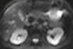
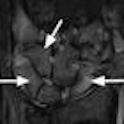
| Images show bone marrow edema in 31-year-old woman with rheumatoid arthritis. Extremity MRI (above) and conventional MRI (below) both show extensive and comparable edematous changes throughout the carpal bones (arrows). Images courtesy of Radiology. |
 |
The readers' image analyses demonstrated "almost perfect agreement between the results on the extremity and conventional MR systems for both readers with regard to bone erosion, synovitis, bone marrow edema, and total scores," the study noted. "Intraclass correlation coefficients were highest for erosion, bone marrow edema, and total scores, but even the synovitis scores demonstrated high agreement between the two systems."
In addition, after each examination, patients were asked to complete a questionnaire to grade their experience regarding claustrophobia, system noise, and difficulty with immobilization. A score of zero was considered as no claustrophobia, noise, or discomfort, whereas a rating of 100 represented maximum difficulty with the examination.
Naraghi and colleagues found that patient feelings of claustrophobia, noise, and difficulty with immobilization were "substantially higher" with the conventional scanner, noting the three patients who failed to complete the study. Overall, 95.8% said they would prefer to have follow-up imaging performed with the extremity scanner.
On the basis of those observations, the researchers concluded that "inflammatory and destructive changes in rheumatoid arthritis are equally well demonstrated with the 1T extremity MR system as with the conventional 1.5T system."
They also added that the 1-tesla extremity system demonstrated "excellent correlation with the 1.5T conventional system, with respect to demonstration of bone marrow edema and synovitis, features that have been problematic for 0.2T extremity systems."
By Wayne Forrest
AuntMinnie.com staff writer
June 8, 2009
Related Reading
MRI with STIR fails to change diagnosis in child abuse cases, May 12, 2009
MRI helps find fractures among elderly female ED patients, April 29, 2009
MRI used to diagnose complex lung infections in children, October 16, 2008
MR parameters divulge depth of acute hamstring injuries, July 13, 2007
MRI keeps pace with rapidly evolving musculoskeletal systems of young athletes, May 20, 2007
Copyright © 2009 AuntMinnie.com