A new study has found that administering intravenous gadolinium does not make a significant difference in MR images of the liver, spleen, or pancreas in a direct comparison of precontrast and postcontrast diffusion-weighted imaging (DWI).
On the other hand, precontrast diffusion-weighted images of the kidney were preferred by a significant margin over postcontrast results.
The findings may allow DWI sequences, which may improve liver lesion detection, to be performed either before or after contrast administration as needed, according to the researchers from Duke University Medical Center in Durham, NC,
In previous research and practice, diffusion-weighted imaging has been shown to improve lesion detection in the liver, peritoneum, and lymph nodes. The technique also has the potential to characterize the lesion and quantitatively assess tumor response to therapy, using apparent diffusion coefficient (ADC) values, noted lead author Dr. Carolyn Lee Wang.
"Early work has shown that we can also characterize chronic pancreatitis and assess chronic hepatic fibrosis, renal disease, and renal artery stenosis using diffusion-weighted imaging and ADC values," she added during her presentation at the recent American Roentgen Ray Society (ARRS) meeting in Boston.
Patient sample
The retrospective study reviewed 50 consecutive patients between August 2007 and February 2008. The patient sample was divided evenly with 25 males and 25 females, with a mean age of 59.4 years.
The patients were referred for contrast-enhanced MR imaging of the abdomen. Clinical indications included staging in known malignancy (18 cases), hepatic disorder or focal liver lesions (17 cases), biliary disorders (10 cases), pancreatitis (two cases), and other conditions (three cases).
Scans were conducted on a 3-tesla MRI (Magnetom Trio with Tim, Siemens Healthcare, Malvern, PA) with a six-channel phased-array coil and embedded spine coil for signal reception. ADC values were calculated automatically by the MRI system and displayed as corresponding ADC maps.
Diffusion-weighted images were acquired prior to and after contrast administration, using single-shot echo-planar imaging with b-values of 50 and 800 s/mm2.
Of the 50 patients, 46 received gadobenate dimeglumine (MultiHance, Bracco Diagnostics, Princeton, NJ), while four patients received gadopentetate dimeglumine (Magnevist, Bayer HealthCare Pharmaceuticals, Wayne, NJ). The volume of contrast was calculated based on patient weight and estimated glomerular filtration rate (eGFR), with a mean of 15 mL.
Image review
Three attending physicians who specialize in abdominal imaging reviewed the pre- and postcontrast diffusion-weighted images, with datasets displayed side-by-side and randomized with the precontrast and postcontrast images to either the left or the right.
The initial dataset of 50 images was then flipped and shown again to the readers, with the entire 100-image dataset randomized prior to presentation. "We used a forced-choice design and asked the attendees which dataset had the best diffusion-weighted appearance for the liver, spleen, pancreas, and kidneys," Wang said. "If an organ was not seen, that data was excluded."
The mean time interval between the precontrast and postcontrast DWI was 16 minutes, with a mean time interval between contrast initiation and the start of postcontrast DWI of 11 minutes. The liver was displayed in all 50 cases, the spleen was visible in 43 cases, the pancreas in 37 cases, and the kidneys in 49 cases.
Readers' evaluations
In reviewing the images, readers preferred precontrast images for the liver 52% of the time, with a p-value of 0.623, which, Wang noted, is not statistically significant. Precontrast images were preferred 49% of the time for the spleen (p = 0.801) and 59% of the time for the pancreas (p = 0.065). "Neither of which was statistically significant," she added.
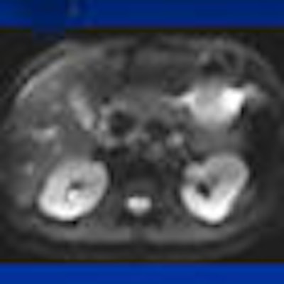
Precontrast images of the kidney were preferred 83% of the time, with a p-value of less than 0.001, which was statistically significant.
 |
| In the precontrast (top three images) and postcontrast (bottom three images) comparison, readers found little difference when viewing the liver and spleen. All images courtesy of Duke University and Dr. Carolyn Lee Wang. |
 |
| Images are representative of the readers' preference of precontrast (top three images) to postcontrast (bottom three images) images of the kidney. |
With those results, the researchers concluded that intravenous gadolinium administration does not make a statistically significant difference in the qualitative appearance of the liver, spleen, or pancreas when comparing precontrast to postcontrast DWI.
"However," Wang added, "the precontrast diffusion-weighted images were preferred in the kidneys, so we recommend performing the sequence before the administration of gadolinium, if the study is tailored toward the assessment of the kidneys."
Study limitations
One limitation of the study is that postcontrast DWI of the kidneys made it "impossible to blind the reader," Wang noted. "However, fortunately, this was not always a very prominent effect nor appreciated. We attempted to minimize this bias by having the readers grade all the other organs before grading the kidneys."
In addition, there was the potential for recall bias. Even though the images were flipped and randomized, a reader may recall having seen a dataset and his or her initial impression. "However, since the intraobserver agreement was not statistically significant, this suggests that this is a minimal bias," she added.
Wang said future research in this area could include evaluating lesion detection and characterization in pre- and postcontrast DWI, as well as evaluating the effects of newer hepatobiliary excretion agents, such as gadoxetate disodium, on postcontrast diffusion-weighted imaging.
By Wayne Forrest
AuntMinnie.com staff writer
May 18, 2009
Related Reading
DWI-MRI predicts pediatric brain trauma outcomes, November 13, 2008
Diffusion-tensor MRI spots subtle changes after mild traumatic brain imaging, March 31, 2008
Diffusion-weighted MRI: The cornerstone of neuroimaging-based stroke studies, June 14, 2007
Diffusion-weighted MR noninvasively monitors uterine fibroid therapies, September 9, 2005
DWI proves best of seven in subacute stroke imaging, May 17, 2004
Copyright © 2009 AuntMinnie.com