Coronary vessel wall imaging is a promising way to depict outward blood vessel modeling when luminal stenosis is absent, potentially improving doctors' ability to evaluate blood vessels with an elevated risk of plaque rupture. A new study from Germany shows that radial k-space sampling is better than Cartesian sampling for examining such vessels in 3D turbo field-echo MRI sequences.
In a small cohort of healthy volunteers, radial k-space sampling produced low motion artifact susceptibility as well as a high signal-to-noise ratio (SNR) using 3-tesla MRI, according to the authors from the University Medical Center Hamburg-Eppendorf in Germany.
The higher field strength successfully compensated for the reduced signal strength inherent in the radial sampling method, explained lead author Dr. Paul Bansmann and colleagues.
"Radial k-space sampling has been shown to be less susceptible to motion artifacts caused by respiratory and cardiac motion," they wrote. "One drawback of the radial k-space sampling method is the reduced -- by a factor of 0.81-- signal-to-noise ratio compared with the corresponding Cartesian images" (American Journal of Roentgenology, January 2007, Vol. 188:1, pp. 70-74).
The authors compared 3D gradient-echo images obtained using radial versus Cartesian k-space sampling -- using a 3-tesla scanner to compensate for the reduced SNR produced with radial sampling.
Coronary MR angiography was performed in nine consecutive volunteers (mean age 24 ± 6 years) weighing an average of 67.2 kg (± 7.6 kg) with an average heart rate of 62.2 bpm ± 4 bpm; all subjects were healthy and in sinus rhythm, with no history of cardiovascular disease. Exams were performed on a 3-tesla scanner (Intera, Philips Medical Systems, Andover, MA) at 30 mtesla/m and a slew rate of 150 mtesla/m/sec, using a six-element phased-array cardiac coil.
A vector ECG was used for cardiac gating. A cine turbo field-echo sequence was applied to analyze the diastolic rest period with a focus on the right coronary artery, and subject-specific trigger delay during the mid-diastolic rest period was identified for each subject to reduce motion artifact, the authors wrote.
To reduce the off-resonance sensitivity of the pencil beam excitation at 3 tesla, k-space turns were reduced to 9 with a beam width of 40 mm, yielding a four-second pulse duration. Fat suppression was applied before the navigator and imaging sequences. Double inversion recovery was applied after the R-wave trigger for blood signal suppression in the right coronary artery. An inversion pulse was followed by a slab-selective pulse to reinvert magnetization in the region of the right coronary artery.
The researchers applied a high-resolution scout image to locate the course of the coronary arteries using a three-point-plan scan tool. Imaging parameters included TR/TE of 13/4.4, flip angle of 30°, turbo field-echo factor of 9, echo-planar imaging factor of 5, acquisition matrix of 256 x 164 x 24, 350 x 280 x 96-mm field-of-view, and sensitivity-encoding (SENSE) acceleration factor of 1.9.
Vessel wall images were acquired using a 3D turbo field-echo sequence (flip angle of 30°, field-of-view of 270 mm, in-plane resolution of 0.7 x 0.7 mm) during free breathing and using prospective navigator gating, with 1.3 oversampling for both sequences.
Cartesian vessel wall imaging of the right coronary artery was performed using TR/TE of 7.8/2.4 and an acquisition matrix of 512 x 358. The scanning duration for the sequence was 616 heartbeats. Radial vessel wall imaging was performed at TR/TE of 8.9/2.3, with a scanning duration of 768 heartbeats. The group acquired 384 radial trajectories in the x-y plane, and both Cartesian and radial images were acquired with six partitions of 2-mm thickness, reconstructed in 12 slices at 1-mm intervals, with an intershot interval of two heartbeats and an acquisition window of 71 msec per shot; SAR was 1.5 W/kg.
 |
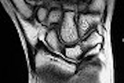
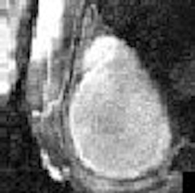
| Reformatted right coronary artery vessel wall images of a healthy 24-year-old man acquired with Cartesian k-space imaging (above) and radial k-space imaging (below). Image republished with permission of the American Society of Roentgenology ©, from AJR 2007; 188:70-74. Paul M. Bansmann, Andrew N. Priest, Kai Muellerleile, Alexander Stork, Gunnar K. Lund, Michael G. Kaul, and Gerhard Adam. |
 |
Bansmann et al used the Soapbubble Tool (Intera, Philips) to analyze visible segments for the measurement of vessel wall, epicardial fat, and luminal blood. The signal was measured in a region of interest and SNR was measured. All images were evaluated by an experienced radiologist and an experienced cardiologist, blinded to sequence parameters and patient information.
According to the results, radial k-space sampling resulted in fewer motion artifacts and improved vessel wall visualization compared to cartesian k-space sampling. Streak artifacts were seen in all radial images, but were easily identified as such and did not affect image quality, the authors wrote.
"Because streak artifacts are around the center of the acquired field-of-view, it is best to avoid having the coronary artery in the exact center of the image," Bansmann and colleagues advised.
They also found a significant difference in the average image quality score for the right coronary artery vessel wall, with an average score of 2.9 for the radial k-space method and an average score of 2.1 for the Cartesian method.
Other measures produced no advantage for either method. No statistically significant difference in vessel length measurement was found between the two methods, and the signal-to-noise and contrast-to-noise ratios at the higher field strength showed no significant difference between radial and Cartesian k-space sampling.
Vessel diameters were equivalent with both methods. However, visual analysis of vessel wall thickness resulted in a larger reported vessel thickness in six of the nine volunteers with Cartesian k-space sampling, and radial k-space sampling yielded a sharper delineation of the vessel wall, the authors wrote.
"MRI of the coronary vessel wall has received considerable attention because a nonstenotic plaque may present a high risk of rupture and stenosis, and may not be visible on luminograms," Bannsman et al wrote. Hence, high-resolution coronary vessel wall scans are needed to detect thickening of plaque within the coronary vessel wall."
Both imaging sequences depicted the right coronary vessel wall adequately, but radial k-space sampling was less susceptible to motion artifacts caused by respiratory and cardiac motion, they wrote. The double inversion-recovery prepulse produced good blood suppression.
"The intrinsic robustness of radial k-space sampling against motion artifacts led to significantly higher image quality scores for radial scans," the authors wrote. "Radial k-space sampling at 3 (tesla) makes it possible to combine low motion artifact susceptibility with high (signal-to-noise ratio)."
By Eric Barnes
AuntMinnie.com staff writer
January 29, 2007
Related Reading
Inflammatory view: Imaging blind to plaque risk, December 7, 2006
MRI discriminates new myocardial infarction from old infarct scar, November 28, 2006
MRA shows good results in detecting coronary stenosis, March 7, 2005
High-field MRI of the brain safely acquired in pacemaker patients, December 15, 2006
Copyright © 2006 AuntMinnie.com