Ultrasound compares favorably with MRI in the evaluation of the posterior tibial tendon (PTT), according to a prospective study presented at the 2002 RSNA meeting in Chicago.
Early recognition of PTT dysfunction is critical in preventing irreversible deformity and disability, said Dr. Krishna Nallamshetty of Thomas Jefferson University in Philadelphia. While MRI has been the mainstay diagnostic modality for PTT pathology, tendon sonography has recently gained in popularity with the advent of high-frequency linear-array transducers and digital imaging, he said.
These advances allow for high-resolution images of the tendon and better depiction of fibrillar architecture, Nallamshetty said. And the results can be correlated directly with the physical examination.
Common PTT injuries seen on ultrasound include a complete PTT tear, a partial tear, and tendinosis, he said. To compare ultrasound and MRI in the evaluation of suspected PTT dysfunction, TJU researchers prospectively studied 22 ankles in 19 female patients, with a mean age of 61.
Evaluated by the same foot and ankle surgeon, all patients had symptoms of PTT dysfunction and more than six weeks of chronic ankle pain. Using a commonly accepted staging system, the surgeon classified five ankles as stage I, 11 as stage II, and six as stage III, Nallamshetty said.
Each of the patients received an MR exam on a 1.5-tesla scanner, and an ultrasound exam using a 10-MHz linear-array transducer on the same day. Three patients underwent bilateral studies.
Each exam was performed and interpreted by experienced radiologists blinded to the results of the other study, and classic clinical findings were utilized as a standard reference, he said.
All stage I ankles were found to be normal on MRI and ultrasound, Nallamshetty said. In the stage II and III cases, MRI and ultrasound exams reported abnormalities in 13 out of 17 ankles, he said. MRI found eight partial tears, one complete tear, and four tendons with tendinosis/tenosynovitis. Ultrasound reported eight partial tears, and five tendons with tendinosis/tenosynovitis.
There were five discordant findings, yielding a concordance rate of 77.3%. In the first three discordant cases, MR found a partial tear, while the ultrasound exam generated a tendinosis finding, he said.
The fourth case resulted in a tendinosis finding on MRI, with a partial tear reported in the ultrasound exam. In the fifth case, MRI diagnosed a complete tear, while the ultrasound study reported a partial tear.
   |
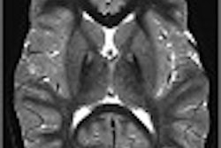
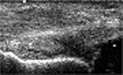
| In this case, ultrasound studies led to a diagnosis of PTT tendinosis with a partial tear (top and bottom), while T1-weighted MR image (center) indicated a complete tear. The patient was treated for a partial tear, however, as the orthopedic surgeon's evaluation of the PTT was more consistent with the ultrasound findings. Images courtesy of Dr. Krishna Nallamshetty. |
None of the discordant readings affected patient management because the clinical symptoms were more consistent with the ultrasound diagnosis, Nallamshetty said. Limitations of the study included a small sample size, an imperfect gold standard, and lack of surgical correlation, he said.
"In this preliminary study, US compared very favorably with MR imaging in the evaluation of PTT, and as a result, we feel that we can use ultrasound as a convenient, faster, and less costly alternative to MR in detecting PTT abnormalities," Nallamshetty said.
By Erik L. RidleyAuntMinnie.com staff writer
February 12, 2003
Related Reading
Pediatric ankle exam predicts need for x-rays, November 1, 2002
Lateral process of the talus fracture plagues snowboarders: a case study, February 24, 2002
Intervention can help or harm in athletic injuries, February 17, 2002
Copyright © 2003 AuntMinnie.com