The cognitive capabilities of humans have long been attributed to a disproportionate enlargement of the brain’s frontal lobe in comparison to a subset of primates. However, this conclusion was based on data that did not include apes, the closest relative to humans, and it did not specifically target a comparison of the hemisphere in front of the central sulcus.
In an article in Nature Neuroscience, Katerina Semendeferi, Ph.D., and colleagues (from the department of anthropology at the University of California, San Diego in La Jolla, CA, and the department of neurology at the University of Iowa in Iowa City) used MRI and 3-D reconstruction techniques to compare the relative size of the frontal cortices in humans, apes of all extant species, and selected monkeys.
"It appears that the traditional notion that disproportionately large frontal lobes and frontal cortices are the hallmark of hominid brain evolution is not supported," the authors wrote (Nature Neuroscience, March 2002, Vol. 5:3, pp. 272-276).
The team obtained MRI scans of the brains of 24 nonhuman primates from the Yerkes Regional Primate Research Center at Emory University in Atlanta. This group was comprised of adults of both sexes of 15 great apes (6 chimpanzees, 3 bonobos, 2 gorillas, and 4 orangutans), 4 lesser apes (gibbons), and 5 monkeys (3 rhesus and 2 cebus).
The primates were imaged on a Gyroscan NT scanner (Philips Medical Systems, Bothell, WA). Axial slices ranged between 1.2 mm and 1.4 mm, with an overlap of 0.6 mm and a field of view of 124-230 cm.
The human group consisted of 10 adults of both sexes who were imaged at the University of Iowa on a 1.5-tesla Signa MRI scanner (GE Medical Systems Waukesha, WI). A protocol of 124 contiguous coronal slices at 1.6-mm thickness in a 256 x 192 matrix and an FOV of 24 cm was used.
"We scanned one human at both locations to make sure that there would be no difference due to the scanner. The animals were sedated, but the humans were not," said Semendeferi in an e-mail to AuntMinnie.com.
 |
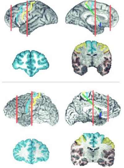
| Three-dimensional reconstruction of the brain of (top) a common chimpanzee and (bottom) a human. The lateral and mesial surfaces of the hemisphere are shown (not to scale). The central and precentral sulci are marked in yellow and cyan, respectively. The posterior end of the frontal cortex is marked by the yellow line (central sulcus). The white segments correspond to connections drawn between discontinuous segments of these sulci. On the mesial surface, the posterior end of the frontal cortex is marked by the solid green line, and the blue line marks the end of the orbitofrontal cortex. The red lines mark the position of the coronal sections through the hemisphere. On the coronal sections, cyan indicates frontal cortices anterior to the precentral sulcus, yellow indicates the cortex of the precentral gyrus, and red the rest of the cortex of the hemisphere. Image courtesy of Katerina Semendeferi, Ph.D., and Nature Neuroscience. |
All MR images were then reconstructed in three dimensions using the Brainvox application (developed by Drs. Antonio and Hanna Damasio, Laboratory of Human Neuroanatomy and Neuroimaging, University of Iowa College of Medicine). The team then calculated the total volume of the hemispheres, the total volume of the cortex of the hemispheres, the volumes of the frontal cortex, the cortex of the precentral gyrus, and the rest of the frontal cortex.
"Although the absolute differences in size of the frontal cortex between humans and other primates were large, the frontal cortex in humans and great apes occupied a similar proportion of the cortex of the cerebral hemispheres," the article stated.
The authors suggested that cognitive abilities previously attributed to an enlarged frontal cortex might be due to differences in the connection of neural circuitry in this sector, or between it and different brain regions. The definitive analysis of the subsectors of the frontal lobe in comparative neuroanatomy could be achieved by a detailed cytoarchitectonic study. But, as the authors point out, due to prohibitive costs and the scarcity of great ape material, such a study is unlikely.
By Jonathan S. BatchelorAuntMinnie.com staff writer
March 28, 2002
Related Reading
Magnetic Resonance Imaging of the Brain and Spine, March 19, 2002
Violent images activate brain region: study, November 18, 2001
Brain scan separates lies from truth, November 16, 2001
MRI study finds that Ecstasy use may lead to axonal brain injury, September 18, 2001
Diffusion MRI allows early assessment of brain tumor treatment, December 27, 2000
Copyright © 2002 AuntMinnie.com


