A new rolling-platform table could make whole-body MR angiography fast and practical for the first time, according to Dr. Mathias Goyen, a radiologist from University Hospital Essen in Germany. AngioSURF's ability to reduce procedure times and increase anatomical coverage has made it popular with both patients and clinicians at the university's MR center, where the table was developed.
In two studies presented at last month's European Congress of Radiology, Goyen and colleagues evaluated AngioSURF's performance, and sought to determine the optimal contrast dose needed to detect pathology in the peripheral vessels. Users of the system were not only able to detect significant pathology, but its longer coverage helped them find unsuspected disease as well.
"Recently, whole-body MR angiography has become possible by combining the contrast-enhanced MRA approach with the latest high-performance gradients," Goyen said. "We are now able to (image) the arteries from the carotids down to the runoff vessels in a single exam. Our preliminary results are quite promising, with sensitivities and specificities for detecting stenoses and occlusions in the mid-80s up to the mid-90s."
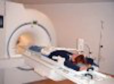
AngioSURF, short for angiographic system for unlimited rolling fields of view, is a manually driven table platform for the acquisition of contrast-enhanced MR angiography.
The device sits atop the patient table of a standard MR scanner, in this case a 1.5-tesla Sonata (Siemens Medical Solutions, Erlangen, Germany). Both a body coil (Siemens CP BodyArray) and a torso surface coil (Siemens CP SpineArray) are integrated into the device to acquire continuous MRA images over as much as 180 cm.
 |
The system is approved for investigational use only; CE Mark approval is pending, according to the manufacturer, MR-Innovation of Essen, Germany, where Dr. Goyen serves as vice president and marketing director.
The first study examined 3 healthy volunteers, as well as 10 patients with angiographically documented peripheral vascular disease. The patients had all undergone digital subtraction angiography (DSA) within the previous 72 hours. Evaluation of the AngioSURF system was based on a total of 214 vascular segments for which DSA was available for comparison.
Each imaging sequence began with a moving vessel scout to plan the 3-D acquisition, followed by a test bolus starting at the level of the first circle, Goyen said.
"We use a biphasic injection scheme with a dosage of 0.2 mmol per kg/body weight of Gd-BOPTA (Bracco Diagnostics, Princeton, NJ)," he said. "And we split the flow rate. For the first half of the volume we inject with a flow rate of 1.3 ml per second. For the second half we reduce the flow rate to 0.7 ml per second to better catch the infrapopliteal arteries."
Imaging was performed using a 3-D fast low-angle shot (FLASH) sequence (TR/TE 2.1/0.7 ms, flip 20º, FOV 400 x 400 mm, matrix 512 x 420 mm, ZIP 512, slab, 160 mm, 64 partitions, effective slice 2.5 mm, RF coils B1, B2, S5, S6). Five contiguous 40-cm volumes with 4-cm overlap 3-D datasets were acquired with 3 cm of overlap, for total craniocaudal coverage of 180 cm. The sequence was acquired over 12 seconds for each of five stations, with 3-second pauses between each station to reposition the table, for a total acquisition time of 72 seconds. AngioSURF's rolling platform enabled easy and precise transition between stations, according to Goyen.
The results were based on 214 vascular segments in which DSA was available for comparison. All patients tolerated the exams well, and all exams were of diagnostic quality, showing complete correlation with DSA findings, Goyen said. For the detection of significant stenoses, the AngioSURF procedures had a sensitivity of 95.3% and specificity of 95.2% compared to DSA.
"Especially for the popliteal arteries, the delineation using the AngioSURF is much better" due to the integration of the body coil and the torso surface coil, he said. SNR (signal-to-noise) and CNR (contrast-to-noise) values are up to three times higher using its integrated torso surface coil rather than a body coil, he said.
 |
"The extended anatomic coverage enabled the diagnosis of a high-grade stenosis of the internal carotid artery that wasn't known before, so we could provide the referring physician with some additional information," Goyen said. The procedure also correctly diagnosed 2 abdominal aortic aneurysms, 1 dissection, and 1 graft status, he said.
 |
"The AngioSURF system allowed the accurate display of arteriovasculature from the supraaortic arteries to the lower extremity vessels," Goyen said. "Due to the extended coverage that's provided, we found additional findings that may be relevant to the treatment and diagnosis of vascular disease."
Finding the right dose
In a second study, the researchers set out to find the optimal contrast dose for MRA with AngioSURF, using the same imaging protocols used in the first study.
"The contrast companies like to say more is better ... but you have to keep in mind that contrast agents are very expensive," Goyen said. "So we have to find some solutions to reduce the amount of contrast that's necessary for different vascular regions."
The purpose of the study was to determine the optimal contrast dosage to provide diagnostic high-resolution whole-body angiograms based on both qualitative and quantitative parameters.
Twelve healthy volunteers each underwent three imaging procedures, with ascending doses of Gd-BOPTA contrast: 0.1, 0.2 and 0.3 mmol per kg of body weight.
According to quantitative results, both SNR and CNR values were significantly higher (p<0.05) with the 0.2 and 0.3-mmol/kg of body weight doses compared to the lowest dose. The results were similar for a qualitative evaluation using a four-point scale, where there was no statistically significant difference between 0.2 and 0.3 mmol/kg of body weight.
"With 0.1 mmol there were actually 63 (vascular) segments that were rated nondiagnostic...whereas with the higher doses all segments were rated as diagnostic," Goyen said. "Our preliminary experience suggests that a dosage of 0.2 is sufficient for diagnostic whole-body MRA, and that increasing the dose to 0.3 [mmol] had no measurable qualitative or quantitative effect."
In response to audience questions, Goyen said whole-body MRA has the potential to change the whole pattern of care for vascular patients, since they can undergo the procedure during the first visit for suspected peripheral vascular disease.
"Angiologists tell us that if we were to do duplex sonography of the whole body it would take about four hours," Goyen said. "There are a lot of patients who get the AngioSURF exam as the first imaging modality. Then we say OK, there are problems over here in the carotids, probably in the renal artery, and then depending on what's the leading pathology, the interventional radiologists and vascular surgeons ... decide what to do."
By Eric BarnesAuntMinnie.com staff writer
April 11, 2001
Click here to post your comments about this story. Please include the headline of the article in your message.
Copyright © 2001 AuntMinnie.com