A group of Japanese researchers has concluded that scanning for a longer period of time is more effective than increasing radiopharmaceutical dose for maintaining PET/CT image quality in large patients.
They found that radiopharmaceutical doses 2.5 times greater than the standard 3.7 MBq per kilogram of body weight did not improve image quality in a PET/CT scanner with high-performance detector electronics and lutetium oxyorthosilicate (LSO) technology.
Led by Dr. Yoko Masuda of Tokyo Women's Medical University School of Medicine and Nagasaki University, the research suggests that lengthening a larger patient's scan time is more effective in maintaining image quality than increasing dose or using other imaging techniques to reduce random and scatter noise. The results are published in the June 2009 issue of the Journal of Nuclear Medicine (Vol. 50:6, pp. 844-848).
The retrospective study first sought to acquire baseline data about image quality in relation to body weight from patients who had undergone clinical PET/CT examinations. The researchers used a standard protocol of injecting an FDG dose of 3.7 MBq/kg and an acquisition time of two minutes for each bed position.
Weight-based patients
The 80 patients (50 men, 30 women) in the baseline study were selected from the researchers' PET center database and categorized into four 20-patient groups based on body weight. All patients had received FDG-PET/CT scans for oncologic conditions between June 2005 and August 2006. As part of the study, they were measured for radioactivity concentrations and signal-to-noise (S/N) ratios in the liver, with comparisons of S/N ratio from the lightest weight group to those in each heavier group.
The researchers then prospectively examined the effects of injected doses or acquisition times on the quality of images in an additional 120 patients based on body weight and matched the protocol for injected doses or acquisition times. The second group of 120 patients (89 men, 31 women) was enrolled prospectively for FDG-PET cancer studies between September 2006 and August 2008. Again, groups of 20 patients were imaged according to the protocol of correction for acquisition time or administered dose.
Images were acquired with an LSO-based PET/CT scanner (Biograph Sensation 16, Siemens Healthcare, Erlangen, Germany). All patients fasted for at least five hours before receiving an intravenous injection of FDG and then rested for approximately 60 minutes.
3D PET data were acquired at eight bed positions from the top of the skull to the middle of the thigh. The CT-based attenuation-corrected FDG-PET images were reconstructed with an ordered-subset expectation maximization (OSEM) algorithm.
Liver uptake
Although liver uptake did not differ significantly among the weight-based patient groups, researchers found that S/N ratio decreased progressively with greater patient weight.
Even with dose administration of 1.4, 1.9, and 2.5 times greater in patient groups 2, 3, and 4 (with the fourth group the heaviest), increased FDG uptake in the liver did not significantly affect S/N ratio compared to the similarly weighted baseline groups. In contrast, the S/N ratio of patient group 4 with the added dose was significantly higher than the S/N ratio of the baseline group with the heaviest patients.
On the other hand, changing the length of time patients were scanned had a major impact on image quality. Group 1 received the standard clinical protocol of two minutes per bed position. In group 2, scanning time was three minutes per bed position, while group 3 received four minutes per bed position and group 4 received five minutes per bed position.
 |
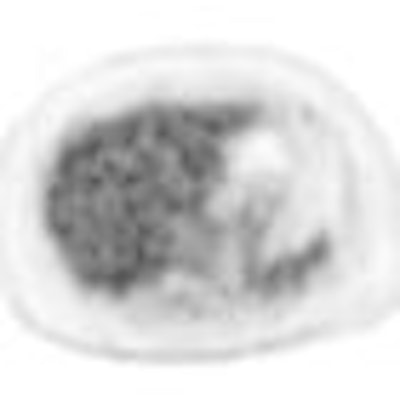
| PET/CT axial liver images show the image quality with average signal-to-noise (S/N) ratio for each patient group. S/N ratios of baseline group 1 (G1, lightest patients), baseline group 4 (G4, heaviest patients), group 4 with higher dose (G4 dose, heaviest group given dose), and group 4 with more scanning time (G4 time, heaviest patients) were 14.8, 8.5, 9.0, and 10.6, respectively. Images courtesy of the Journal of Nuclear Medicine. |
The researchers found that the S/N ratio of group 4 with the standardized scanning protocol was 8.5, whereas the S/N ratio of group 4 with a scanning time of five minutes per bed position was 10.6, a difference that was statistically significant.
Masuda and colleagues concluded that their major finding was that an "extended acquisition time effectively maintained the quality of FDG-PET/CT images of overweight patients. In contrast, an increased dose of up to 2.5-fold higher than 3.7 MBq per kilogram of body weight did not improve image quality. Our findings suggest that only scanning for prolonged periods can maintain the quality of images of heavier patients."
They also determined that liver uptake was consistent among patients administered the same dose per kilogram of body weight, and image quality became degraded as body weight increased. Additional increases in dose did not affect image quality.
The authors cited several limitations of the study, including the fact that image quality at baseline was not compared with image quality when the time or dose was adjusted in the same patients. They noted that S/N ratio of all the patient groups "tended to improve after the time adjustment, but reached statistical significance only in the heaviest group."
By Wayne Forrest
AuntMinnie.com staff writer
June 10, 2009
Related Reading
PET/CT confirms presence of brown fat in adults, April 22, 2009
PET/CT shows early chemo results, April 16, 2009
FDG-PET/CT helps predict osteosarcoma survival, March 12, 2009
FDG-PET aids in detecting atherosclerosis activity, February 23, 2009
FDG-PET/CT recommended for staging Hodgkin's disease, December 19, 2008
Copyright © 2009 AuntMinnie.com