Researchers at Children's Hospital Boston and Harvard Medical School in Boston have found that they can get better image quality and lower radiation dose in pediatric SPECT renal studies by using a data reconstruction technique based on ordered subset expectation maximization with 3D resolution recovery (OSEM-3D).
The study compared OSEM-3D to the standard SPECT reconstruction technique, filtered back projection (FBP), in reconstructing technetium-99m (Tc-99m) dimercaptosuccinic acid (DMSA) renal SPECT data. The research was led by Dr. Niall Sheehy and was published in a March 20 online version of Radiology.
Although OSEM-3D reconstruction is known to improve image quality, the study noted that the technique has not been implemented to a great degree in clinical practice, and has not previously shown definitively whether the enhanced image quality significantly reduces radiopharmaceutical activity or scan time.
At Children's Hospital, the nuclear medicine department routinely uses a dual-detector gamma camera, with each detector rotating a full 360° around a patient. Data reconstruction is performed using an FBP algorithm.
Patient review
The researchers retrospectively reviewed studies conducted on 50 patients: 14 males between 1 and 22 years of age (mean age, 10 years) and 36 females between 1 and 20 years (mean age, 8 years). The study included a total of 98 kidneys.
All 50 patients had been referred for renal Tc-99m DMSA SPECT imaging between January 2007 and April 2007. The patients intravenously received 1.85 MBq of Tc-99m DMSA per kg of body weight, with a dose range of 7.4 to 111.0 MBq. SPECT imaging took place approximately four hours after injection.
The scans were all conducted on the same gamma camera (e.cam, Siemens Healthcare, Malvern, PA) equipped with ultrahigh-spatial-resolution collimators.
The researchers noted that because only a single detector was used for the OSEM-3D reconstruction, they were able to generate datasets "with 50% fewer counts than were needed for the FBP reconstruction without altering our clinical imaging protocol or scanning the patient again."
Two nuclear medicine physicians blinded to patient histories reviewed the results after being presented with the reconstructed SPECT datasets. They analyzed the images using three-view SPECT display (transverse, sagittal, coronal) images and rotating maximum intensity projection (MIP) images.
Image evaluation
The reviewers evaluated the images for overall quality with a five-point scale, ranging from grade 1, indicating a nondiagnostic image, to grade 5, indicating an excellent quality image with "crisp definition" of the cortex and the renal septa.
The results showed that "although the OSEM-3D images were reconstructed with 50% fewer gamma photon counts than were the FBP images, the image quality of the OSEM-3D studies (mean score, 4.3 ± 0.7 [standard deviation]) was superior to that of the FBP studies (mean score, 3.5 ± 0.9) (p < 0.001)," the authors wrote.
 |
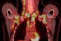
| The images show maximum intensity projection Tc-99m DMSA renal SPECT of a four-year-old girl with multiple urinary tract infections and duplex left collecting system. A voiding cystogram revealed a duplex system on the left, with vesicoureteral reflux in left lower pole. Both the full-count FBP (above) and half-count OSEM-3D SPECT (below) images show loss of left renal parenchymal function in left lower pole, with focal cortical defects, but the results are clearer in the OSEM-3D image below. Images courtesy of Radiology and Children's Hospital Boston. |
 |
The reviewers rated the mean renal cortical defect scores (on a 15-point scale) for OSEM-3D at 14.3 ± 1.4 and FBP as 14.4 ± 1.4. Both methods yielded a median score of 15, with a range of nine to 15, which indicates that the majority of the cases had normal findings.
OSEM-3D's performance
The authors also noted that both techniques yielded identical findings for 94 of the 98 kidneys evaluated: 19 kidneys had cortical defects, and 75 had no cortical defects. Four kidneys showed cortical defects at OSEM-3D but not on FBP images.
The authors concluded that the study "established the feasibility of reducing the total administered radiopharmaceutical activity in pediatric patients by at least a factor of two without sacrificing image quality."
In addition, with the use of standard radiopharmaceutical activity levels, scanning time "can be reduced by half to result in increased patient comfort and/or a minimized need for sedation."
Depending on the clinical circumstance, use of OSEM-3D also may enable nuclear medicine practitioners to reduce radiation dose and shorten imaging time. That capability could help customize nuclear medicine exams for the individual patient and improve overall disease management and therapeutic outcomes in the future.
By Wayne Forrest
AuntMinnie.com staff writer
May 5, 2009
Related Reading
SPR news: Rads must take lead in reducing pediatric CT dose, April 23, 2009
Study: Radiologists dial back on pediatric CT settings, October 4, 2008
CT experts grapple with rising concerns about radiation dose, May 14, 2008
FBP and AW-OSEM square off in 2D/3D cardiac PET, July 2, 2004
Copyright © 2009 AuntMinnie.com