Radiographically dense breasts continue to pose a challenge to women's imaging specialists. The alternative imaging options that have been proposed include computed radiography for mammography, as well as ultrasound, MRI, and scintimammography. A study in the latest issue of the Journal of Nuclear Medicine has suggested taking the latter modality one step further, with a dedicated breast camera.
"The value of scintimammography as an adjuvant to standard screening modalities is in the early detection of breast carcinoma. Scintigraphy may be appropriate for the subset of women whose breasts are difficult to examine by conventional means," wrote Dr. Leonard Coover and colleagues. However, "the sensitivity of scintimammography with a standard gamma camera is dependent on lesion size" (JNM, April 2004, Vol. 45:4, pp. 553-558).
Previous reports have put the sensitivity of standard scintimammography for nonpalpable tumors at about 55%, added Coover, who is from the Hamot Medical Center in Erie, PA. Co-author Dr. Gina Caravaglia is from Loyola University in Chicago, while Phyllis Kuhn, Ph.D., is from the Lake Erie Research Institute, Erie, PA.
Using a dedicated breast camera -- one that can be adapted to fit on most upright mammography machines (replacing the bucky) -- allowed for breast compression during imaging, which facilitates lesion detection.
The researchers tested their technique on 37 women with BI-RADS 1 or 2 findings on mammography. These women also had BI-RADS patterns of heterogeneously dense or extremely dense breast tissue.
The camera (LumaGEM, Gamma Medica, Northridge, CA) had a compact detector head (15 x 12.5 x 8.5 cm) and weighed 10 kg, the authors reported. During image acquisition, the breast was compressed with the tissue spread over the camera face, which reduced background density and improved lesion contrast.
"The mammography gantry allowed flexibility in positioning," they said. "If a lesion was suspected in an area that was not close to the camera, the gantry was repositioned and additional views were acquired."
The women were injected with 740 MBq of Tc-99m sestamibi (Bristol-Myers Squibb Medical Imaging, N. Billerica, MA), with imaging starting 10 minutes later. A lateral view of each breast was acquired, along with an anterior view. An hour later, after another radiocontrast injection, each breast was imaged in the caudal and lateral oblique views.
 |
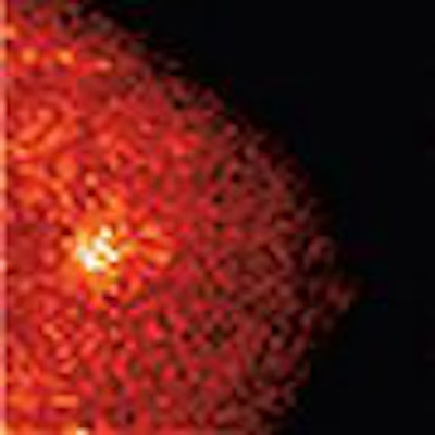
| Patient 1 with infiltrating lobular carcinoma. Mediolateral-oblique mammogram failed to demonstrate a nonpalpable lesion that was obscured by dense fibroglandular tissue. The circled, loosely grouped calcifications were anterior to the tumor and had been stable for years. The lesion was interpreted as BI-RADS category 2. Reprinted by permission of the Society of Nuclear Medicine from: Coover LR, Carvaglia G, Kuhn P, Scintimammography with Dedicated Breast Camera Detects and Localizes Occult Carcinoma, J Nucl Med 2004; 45:553-558. |
Two reviewers interpreted the images independently, with findings deemed positive or negative based on the identification of focal, increased sestamibi uptake with definable borders. Imaging results were compared to biopsy results.
The dedicated breast camera yielded positive results in 13.5% (five) of the 37 women. The biopsy results in three of these patients showed infiltrating lobular carcinoma, ductal carcinoma in situ, and infiltrating lobular carcinoma, respectively.
In comparison, imaging with a standard gamma camera showed positive results in 8.1% of the patients (three out of 37). Only the infiltrating lobular carcinoma was found on the standard gamma camera scan.
 |
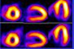
| Scintimammogram of patient 1 obtained with a dedicated breast camera. Better signal-to-noise ratio allowed lesion detection. Biopsy revealed infiltrating lobular carcinoma. Reprinted by permission of the Society of Nuclear Medicine from: Coover LR, Carvaglia G, Kuhn P, Scintimammography with Dedicated Breast Camera Detects and Localizes Occult Carcinoma, J Nucl Med 2004; 45:553-558. |
"In patient 1, the mammographic results were negative...these three carcinomas were undetectable by clinical breast examination or by mammography, even on retrospective review," the authors said.
In addition to improved cancer detection in women with dense breasts, the authors suggested that scintimammography may also have value in women with fibrocystic changes, implants, or scarring from previous surgery or radiation treatment. They concluded that a study with a larger patient population was in order to further validate their promising results.
Other investigators have reported similarly positive results. In a talk at the 2003 RSNA meeting in Chicago, Dr. Rachel Brem and colleagues from George Washington University Medical Center in Washington, DC, evaluated high-resolution scintimammography in high-risk asymptomatic women with dense breasts. They scanned 180 women with BI-RADS 1 or 2 mammograms using 20 mCi Tc-99m sestamibi on a Dilon 6800 gamma camera (Dilon Technologies, Newport News, VA).
Of these 180 women, the scintimammograms detected 13.3% who had radiotracer uptake. And of those who underwent biopsy, 15% had a diagnosis of malignancy, they reported. In total, unsuspected breast cancer was found in 1.7% of the study population.
More recently, a group from Pusan National University in South Korea established optimal visual grades for the detection of primary breast cancer with scintimammography. They looked at 520 patients with breast cancer (370 malignant; 150 benign).
Based on various lesion-to-non-lesion cutoff points, the sensitivity of scintimammography for detecting malignant lesions ranged from 77% to 86%, they reported. They also found that breast imaging with a gamma camera may be at its best for finding tumors that are 3 cm or smaller (Breast Cancer Research and Treatment, January 2004, Vol. 83:2, pp. 129-138).
By Shalmali PalAuntMinnie.com staff writer
April 20, 2004
Related Reading
ACRIN updates multiple breast cancer trials, April 15, 2004
CR mammography increases visibility of lesions in dense breasts, study finds, April 9, 2004
Mammography affected by dense breasts in postmenopausal women, March 17, 2004
New scanner may find cancer earlier than mammogram, December 5, 2003
Women with genetic history of breast cancer benefit from MR screening, November 30, 2003
New camera may improve scintimammography, August 6, 2002
Copyright © 2004 AuntMinnie.com