In a world with few noninvasive options for detecting prostate cancer, a short-half-life radiotracer could be king. Today Japanese researchers report unambiguous success with a new PET technique for diagnosing prostate cancer in its early stages.
A study in this month's Journal of Nuclear Medicine puts 11C-acetate on a very short list of radiopharmaceuticals that could prove useful for imaging slow-growing prostate cancers, most of which have responded poorly to the more readily available 18fluorodeoxyglucose (FDG). 18F-choline has also shown promise for detecting prostate cancer in studies of mice and men.
The current research, by Drs. Nobuyuki Oyama, Hironobu Akino, and colleagues from the Fukui Medical University, found that 11C-acetate PET detected 100% of biopsy-proven prostate cancers, compared with 83% sensitivity with FDG-PET (Journal of Nuclear Medicine, Vol. 43:2, pp. 181-186).
"The success of (FDG) PET in many cancers has led to the evaluation of this pharmaceutical for use with prostate cancer, the most commonly diagnosed cancer in men and the second leading cause of cancer death in men older than 40 years in the U.S.," the authors wrote.
Unfortunately, FDG-PET cannot reliably detect prostate cancer, having spotted just 50% to 80% of the adenocarcinomas in previous studies. The authors attribute FDG's lackluster showing to the slow glucose metabolism of most prostate cancers, and to other factors such as significant excretion of the tracer into the urinary bladder.
There are circumstances in which FDG has shown a high sensitivity for prostate cancer, Oyama and colleagues wrote, "but only when there is a high tumor viability, such as a high histologic grade, a high clinical stage, or a patient with a high serum prostate-specific antigen (PSA) value."
Moreover, anatomical imaging modalities are of limited usefulness in diagnosing prostate cancer because anatomic changes are not always present in the early stages of disease.
Twenty-two patients (ages 52-85, median age 72.0) participated in the study. All had biopsy-proven prostate cancer that had not yet been treated at the time of imaging. According to the staging criteria of the TNM Classification of Malignant Tumors, 4 patients had stage T1/T2 cancer; 4 had stage T3/T4, and 10 had stage N(+)/M(+).
All patients underwent C-acetate PET following injection of 740 MBq of 11C-acetate. After fasting for at least four hours, 18 of the patients also underwent 18-F FDG-PET imaging within 1 week of the C-acetate exam. For coregistration purposes, the patients also underwent pelvic CT or MRI, as well as bone scintigraphy when warranted.
 |
| PET images of prostate obtained using 18-F-FDG and 11-C-acetate compared with CT image from 71-year-old man with well-differentiated (Gleason sum 2) adenocarcinoma of prostate. (A) CT shows only minimal enlargement of prostate. (B) 18-F FDG-PET shows low uptake in prostate, with SUV of 1.97. (C) 11-C acetate PET shows high uptake in primary prostate cancer lesion, with SUV of 6.41 Note: pr=prostate. Image and caption republished with permission of the Society of Nuclear Medicine from Oyama et al, February 2002, Vol. 43:3, pp. 181-186. |
The data were analyzed by drawing a circular region of interest around the area of the highest radiotracer uptake on transaxial PET images, at locations corresponding to the anatomic location of the prostate shown on reference CT or MRI images. The standardized uptake value (SUV) was calculated for each patient, and the mean SUV value was used to indicate C-acetate and 18-F FDG uptake in the region.
According to the results, adenocarcinoma corresponded with C-acetate uptake, with SUVs ranging from 3.27 to 9.87. In contrast, 18-F FDG uptake ranged from 1.97 to 6.34. Visual inspection showed C-acetate accumulation in all 22 patients, while FDG was found in 15 of 18 patients. (One such patient could not be evaluated due to high FDG accumulation in the bladder.)
"Of the five patients who had lymph node metastases, C-acetate PET showed all to have high intrapelvic accumulations corresponding to metastatic sites," the authors wrote, in contrast to FDG accumulations in 2 of 5 patients who underwent that examination.
In addition, bone scintigraphy revealed bone metastases in 7 of the patients, corresponding to high localized 11C-acetate uptake in 6 of the patients, compared with only 4 for FDG. An evaluation of tumor viability showed no correlation between the Gleason sum and uptake of either C-acetate or FDG.
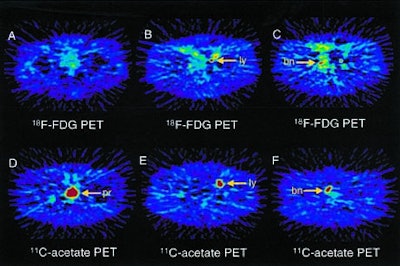
 |
| PET images of prostate, lymph node, and bone metastases obtained using 18-F FDG and 11-C-acetate from 73-year-old man with poorly differentiated (Gleason sum 7) adenocarcinoma of prostate. (A) 18-F FDG-PET shows low uptake in prostate, with SUV of 2.87. (B and C) 18-F FDG uptake was shown in left iliac lymph node metastatic lesion (B) and right pubic bone metastatic lesion (C). (D-F) 11-C-acetate PET shows high uptake in prostate (D), with SUV of 5.45; in left iliac lymph node metastatic lesion (E); and in right pubic bone metastatic lesion (F). Note: bn=bone; ly=lymph node; pr=prostate. Image and caption republished with permission of the Society of Nuclear Medicine from Oyama et al, February 2002, Vol. 43:3, pp. 181-186. |
The authors cited a previous study by Yoshimoto et al for insight into 11C-acetate's affinity for malignant cells. The agent's "accumulation in tumor cells was shown to be caused by enhanced lipid synthesis," Oyama et al wrote. "Given the highly active basal lipid metabolism associated with the cell membrane because of tumor growth, 11C-acetate may be an important probe of this anabolic pathway of metabolism in cancer tissue" (Nuclear Medicine and Biology 2001, Vol. 28, pp. 117-122).
And in a study at the University of Michigan in Ann Arbor, 11C-acetate PET showed high sensitivity for detecting both primary and nodal metastatic prostate cancer lesions (Journal of Nuclear Medicine 1999, Vol 40 [suppl], 60 p).
"11C-acetate is a promising tracer for imaging prostate cancer and its metastases," Oyama et al concluded. "Further studies using C-acetate PET with a larger number of patients are needed to determine its ultimate clinical utility."
In an e-mail to AuntMinnie.com, Dr. Edward Coleman from Duke University in Durham, NC, agreed that 11C-acetate shows great promise -- and needs more research.
"The article by Oyama et al is further documentation that PET imaging of prostate cancer is feasible using 11C-acetate, and that acetate imaging is superior to FDG imaging in prostate cancer," Coleman said, adding that such preliminary studies are laying the foundation for further documentation of the tracer's utility. At the same time, he believes the agent's short half-life could be a problem.
"Although the carbon-11 label with its half-life of 20 minutes is a potential limitation to its widespread utilization, the agent could be made available to multiple centers if the imaging was shown to have a major impact on the management of these patients," Coleman said. "A fluorine-18-labeled agent would have the advantage of the 110-minute half-life, which does permit wide distribution."
Coleman and colleagues are currently evaluating 18F fluorocholine (FCH) for imaging prostate cancer (Cancer Research, January 2000, Vol 61, pp. 110-117). It's too early to tell which agent will ultimately work better, he said.
By Eric Barnes
AuntMinnie.com staff writer
February 15, 2002
Related Reading
Croatia has Europe's greatest increase in cancer mortality, October 12, 2001
Nordion begins yttrium-90 production, August 27, 2001
Auto co-registration of MR, SPECT images could avoid prostatectomy, June 6, 2000
Nuclear medicine researchers seek better prostate imaging with new gamma probe, July 6, 2000
Copyright © 2002 AuntMinnie.com