Osteopenia is the key to separating acute hip fractures from other bone conditions that can seem remarkably similar in the emergency room. With the graying of the population, it’s likely that healthcare professionals will be seeing more of these potentially confusing cases, a hypothesis that has borne itself out in Rochester, NY, according to Dr. Johnny Monu.
"In my neck of the woods, Rochester is known as the snow capital of the world. Once you have snow on the ground, you have elderly people falling all over the place," said Monu, an assistant professor of radiology at the University of Rochester. At last month's European Congress of Radiology in Vienna, Monu shared his experiences in treating these patients.
In his first presentation, Monu discussed collar osteophytes as the cause of false-positive bone scans for hip fractures. At his institution, nuclear medicine scans are ordered when plain films are negative or equivocal, he explained. While the causes of false-negative scans have been documented (bone marrow edema, nontraumatic avascular necrosis), the causes of false-positive scans are not as well known, he said.
Monu began by identifying patients from the hospital database who were positive for hip fracture on the bone scans, but negative on MRI. These patients also had equivocal x-rays initially and follow-up CT scans.
"The MRI scans were done within 72 hours of the bone scans and plain films. MRI and the CTs were done as follow-up especially when there was strong dichotomy between the plain film and bone scan readings, or if the bone scans were equivocal," Monu told AuntMinnie.com. "The multiple studies reflect the various zones of comfort and thinking of the clinicians looking after these patients."
Bone scans were acquired on dual-headed cameras, three hours after intravenous administration of 25-30 mCu of 99technetium methylene diphosphonate (Tc-MDP).
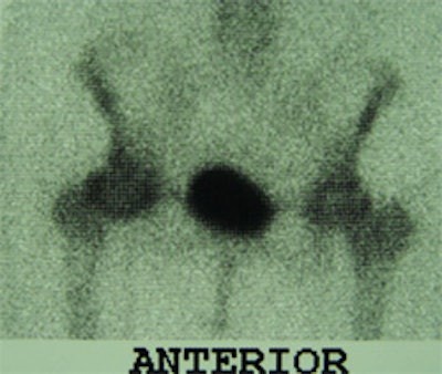
Reviewing the records of these five patients, Monu said he found that all of them had a ring of osteophytes around the femoral neck, which can mimic a fracture.
This is non-precaptioned photo 1

"Even if you used SPECT, because you are taking a slice around the femoral neck, you will get a linear appearance of a fracture," he said. "None of these patients had a fracture. All of them had osteoarthritis. One went on to have hip replacement surgery and the others were successfully treated with NSAIDS [nonsteroidal anti-inflammatory drugs]."
Osteopenia, or low bone density, is a major factor in determining whether a patient requires imaging beyond the initial x-ray, Monu said.
"If you don’t have osteopenia, and you don’t have a history that would suggest fracture, then you have to look elsewhere. A patient with a [true] femoral neck fracture requires hip surgery, and that comes with risks," he said.
In a second presentation, Monu elaborated on the role of MRI to examine potential hip-fracture patients, and the pattern of injuries that may occur.
This is non-precaptioned photo 2
"Patients will present with suspicion of hip fracture, and the plain films are lousy," Monu said, noting that the patients may have other unsuspected abnormalities. "We wanted to see if MR could be used to explain some of the patient symptoms. Every patient had T1-weighted imaging and some form of T2-weighted imaging."
Sorting through four years of hospital records, Monu found 53 patients who had been examined for suspected hip fractures. Thirty-one patients had bone trauma either in the femur or in the ipsilateral innominate bone, while the remaining 22 had no bone abnormalities. More than half of the patients had joint effusions, Monu said.
In 34 cases, an abnormal signal was seen in the obturator externus muscle. Other muscles that had an abnormal signal indicating edema included the abductor brevis (21%), the obturator internus (15%), the gluteus medius (15%), and the adductor magnus (15%). In 13% of the cases, there was evidence of trauma to the gluteus maximus and adductor longus.
"We saw that the majority of these patients had some form of bone injury," Monu said "We also found that in patients with soft-tissue injuries, almost all of the muscles in the hip area were involved."
By Shalmali PalAuntMinnie.com staff writer
April 5, 2001
Related Reading
Hip arthroplasty should not be delayed until radiographic changes are severe, March 26, 2001
MRI defines source of hip pain when plain film fails to produce results, March 3, 2001
Click here to post your comments about this story. Please include the headline of the article in your message.
Copyright © 2001 AuntMinnie.com