Fine-tuning the diagnosis and treatment of breast malignancies remains the paramount concern of women's healthcare. But gynecologic malignancies are also a major problem: worldwide, cervical cancer affects more than 400,000 women, while ovarian cancer accounts for 4% of all cancers in women, according to the Society of Gynecologic Oncologists.
In the U.S, endometrial cancer is the most common cancer of the female reproductive tract. Close to 36,000 women were diagnosed with the disease in 2000, and approximately 6,500 women died from it (National Foundation for Cancer Research).
International investigators are turning to PET imaging to aid in the diagnosis and management of gynecologic tumors. Thus far, they’ve reported positive results with their modalities of choice -- FDG-PET, PET/CT, and choline PET.
Dialing down radioactivity
In one study, Japanese researchers compared 11C-choline PET to FDG-PET for the detection and localization of cancer. Past studies have shown that malignant tumors in the pelvis can be problematic on FDG-PET because of tracer radioactivity spilling into the bladder.
"Because 11C-choline shows only minimal radioactivity in the urinary tract and bladder of fasting individuals, prostate tumor can be visualized more clearly...this fact implies that 11C-choline may be feasible for the detection of pelvic tumors; however, little (is) known about the interpretation of gynecologic tumors by 11C-choline PET," wrote lead author Dr. Tatsuo Torizuka, Ph.D., in the Journal of Nuclear Medicine. Torizuka and colleagues are from the Hamamatsu Medical Center in Hamakita (July 2003, Vol. 44:7, pp. 1051-1056).
For this study, 21 patients with gynecologic tumors underwent choline PET and FDG imaging on a whole-body PET scanner (SHR22000, Hamamatsu Photonics, Hamamatsu City, Japan). Static emission scanning was performed one hour after intravenous administration of 400-500 MBq of 18F-FDG. Dynamic emission scanning was performed after IV administration of 11C-choline (500-600 MBq).
Of the 21 women, 18 underwent PET examinations before surgery. Based on histology, uterine corpus cancer was the primary tumor in 11 cases; uterine cervical cancer was the diagnosis in five cases; and there was one case each of ovarian cancer and pelvic inflammatory disease.
For the detection of uterine corpus cancer, the authors reported that both choline PET and FDG-PET were true-positive in nine cases. In patients with uterine cervical cancer, choline PET was deemed successful because of low urinary radioactivity. In comparison, tumor visualization on FDG-PET was partly hampered by intense bladder activity.
 |
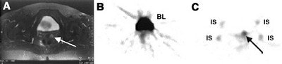
| Transaxial T2-weighted MR image (A) shows a small hyperintense lesion in the uterine cervix (arrow). In FDG-PET scan (B), the tumor is obscured by high bladder activity. The choline PET image shows intense tumor uptake (arrow), corresponding to cervical cancer. Reprinted by permission of the Society of Nuclear Medicine from: Tatsuo T et al. Imaging of Gynecologic Tumors: Comparison of 11C-Choline PET with 18-F FDG-PET. J Nucl Med 2003; 44:1051-1056, Figure 3. |
In three patients with recurrent ovarian cancer, imaging with both tracers showed true-positive results by detecting paraaortic lymph node metastasis in the abdomen. However, neither method was able to detect cystic recurrent tumor and microscopic disseminating disease.
Overall, findings from both methods were true-positive in 15 patients, false-negative in three cases, and false-positive in one patient. Choline PET showed a higher sensitivity (85%) than FDG-PET (75%) for tumor detection. But the mean standardized uptake value (SUV) was lower (4.61) for choline PET than it was for FDG-PET (9.14) in the five patients with true-positive results.
"11C-choline PET can clearly detect primary tumors because of low urinary activity; however, variable physiologic accumulation of 11C-choline in the intestine may be a problem," the authors concluded.
One major benefit of choline PET is a shorter waiting period between tracer injection and imaging: 60 minutes or longer with FDG compared to 3-5 minutes for choline.
PET/CT for better staging
In another study, Israeli investigators from the Rambam Medical Center and the Israel Institute of Technology, both in Haifa, tested hybrid PET/CT imaging with FDG for managing patients with gynecologic malignancies, including cervical and ovarian cancer.
Cervical carcinoma is the most common gynecological malignancy, said Dr. Ora Israel during her presentation at the 2003 Society of Nuclear Medicine conference in New Orleans. However, using CT alone to diagnose cervical cancer can be misleading, as the modality relies on the detection of abnormal or enlarged organs, which may not tell the whole story.
For this study, 57 patients were included, of which the majority (38) had cervical cancer. Thirteen had ovarian cancer and six had cancer of the endometrium. Of these 57 women, 17 were evaluated for staging at the time of diagnosis while recurrence was suspected in the remaining 40. Overall, 86 suspicious lesions were evaluated.
After the injection of 10-15 mCi FDG, 20 patients were evaluated using a SPECT camera in high-energy mode with a CT tomogram module (Millennium VG and Hawkeye, GE Medical Systems, Waukesha, WI), while 37 patients were imaged with a dedicated PET/CT unit. The PET and CT images were first interpreted independently, followed by an analysis of the fused images. The latter was assessed for its additional value and impact on patient management, Israel said. All imaging results were correlated to histologic, radiologic, and clinical follow-up data.
According to the results, PET/CT improves image interpretation over PET and/or CT in 56% of the lesions and in 51% of the patients, and it also provides better localization. It helped clarify abnormal or equivocal FDG uptake, which resulted in retrospective lesion detection.
Overall, PET/CT exams led to a change in management for 30% of patients, mostly those with cervical cancer. Surgery was canceled and alternative treatments -- radiotherapy and chemotherapy -- were instituted in 10 patients. Other changes included alteration of the radiation field, redefinition of a surgical approach, and a switch from curative to palliative therapy.
Israel and colleagues concluded that PET/CT had value in more than half of their patients, and that the modality may be key to avoiding the morbidity and expenses associated with incorrect or unnecessary surgery.
Recurrent cancer
A second presentation at the SNM meeting, this one from Johns Hopkins Medical Institutions in Baltimore, compared PET/CT to PET for detecting residual disease in patients with ovarian cancer, and found the hybrid modality to have a slight edge in specificity.
In this retrospective study, the FDG-PET and PET/CT scans (Discovery LS, GE Medical Systems) of 46 patients with a history of ovarian cancer were evaluated, said Dr. Christian Cohade. FDG-PET images with CT-based attenuation correction and PET/CT images were interpreted by one observer who scored each identified lesion on a five-point scale ranging from definitely benign to definitely malignant. Findings were correlated with pathology, additional imaging, serum markers, or clinical follow-up. Cohade pointed out that only macroscopic disease, greater than or equal to 1 cm, was considered significant for positive surgery.
FDG-PET detected 156 lesions (grades 3 and 4) while PET/CT pinpointed 159. The number of equivocal lesions was reduced from 17 to 9 based on PET/CT results. For the detection of macroscopic disease, the sensitivity of PET was 80%, the specificity was 81.3%, and the accuracy was 80.4%.
In comparison, the sensitivity of PET/CT was 80%, the specificity was 87.5%, and the accuracy was 82.6%. The agreement between the two modalities had a 95% confidence interval. PET and PET/CT results were discordant in three cases. In one patient, focal ureteral activity was misinterpreted as recurrence on PET and as urinary activity by PET/CT.
"PET/CT was able to reduce the number of equivocal lesions by 47% in comparison to PET," Cohade said. "With PET/CT, we were able to improve specificity."
By Shalmali PalAuntMinnie.com staff writer
August 4, 2003
Related Reading
Obesity may double risk of cervical adenocarcinoma, July 23, 2003
Chemotherapy tops radiation in endometrial cancer, but with high toxicity, June 4, 2003
Endometrial cancer can develop after radiation for cervical carcinoma, May 13, 2003
Cryoablation device provides durable results for uterine bleeding, April 22, 2003
New reports address prostate cancer staging, therapy, March 24, 2003
Copyright © 2003 AuntMinnie.com